
Watch Webinar
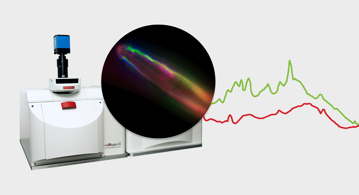
A groundbreaking technique that combines submicron infrared spectroscopy with fluorescence detection is revolutionizing chemical imaging at the microscale. In the webinar, Dr. Craig Prater presented Widefield Super-Resolution Infrared Spectroscopy using Fluorescence-Detected Photothermal IR (FL-PTIR), a novel approach that significantly enhances the sensitivity and speed of traditional photothermal infrared spectroscopy.
The technique, developed independently by multiple research groups including Dr Prater’s team at Photothermal Spectroscopy Corp and recently commercialized, leverages the temperature dependence of fluorescence emission to detect infrared absorption.
When pulsed infrared light is absorbed by a sample, it creates localized temperature increases that result in detectable decreases in fluorescence emission. This approach delivers approximately 100 times greater sensitivity than conventional photothermal methods, which rely on measuring small changes in refractive index.
What makes FL-PTIR particularly valuable for biological applications is its ability to utilize native autofluorescence present in many biological samples, eliminating the need for external fluorescent dyes. Dr Prater demonstrated this capability on various specimens, including micro-algae, plant tissues, mammalian samples, and diatoms, achieving sub-300 nanometer spatial resolution.
The system works by illuminating samples with a pulsed infrared beam while simultaneously exciting fluorescence with visible light. A camera captures fluorescence images with and without IR pulses, and the difference between these images creates an infrared absorption map. This widefield detection approach allows simultaneous measurement at over 260,000 sample points, making it approximately 50 times faster than single-point techniques.
Several impressive applications were showcased during Dr Prater’s presentation. These included:
- Chemical differentiation of polymer microspheres with identical fluorescent labels but different infrared absorption profiles
- Detailed imaging of collagen samples demonstrating molecular orientation effects
- Analysis of kidney tissue heterogeneity using principal component analysis and fuzzy c-means clustering
- High-resolution chemical mapping of diatom silica structures with distinctions between different silicon-oxygen bonding arrangements
- Dynamic imaging of living microorganisms in aqueous environments
The technique’s ability to conduct hyperspectral imaging—collecting complete infrared spectra across an entire sample—in minutes rather than hours represents a significant advancement. This enables researchers to perform sophisticated multivariate analyses that reveal subtle chemical variations across samples.
For samples that don’t exhibit natural fluorescence, Dr Prater noted that conventional photothermal approaches remain available on the same platform. Additionally, for samples sensitive to laser damage, alternative low-power detection methods using avalanche photodiodes can be employed.
This innovation bridges the gap between conventional infrared spectroscopy’s chemical specificity and fluorescence microscopy’s high spatial resolution and sensitivity. By enabling rapid chemical imaging with sub-diffraction spatial resolution in both labeled and unlabeled biological specimens, FL-PTIR opens new possibilities for studying biochemical processes at previously inaccessible scales and speeds.
