
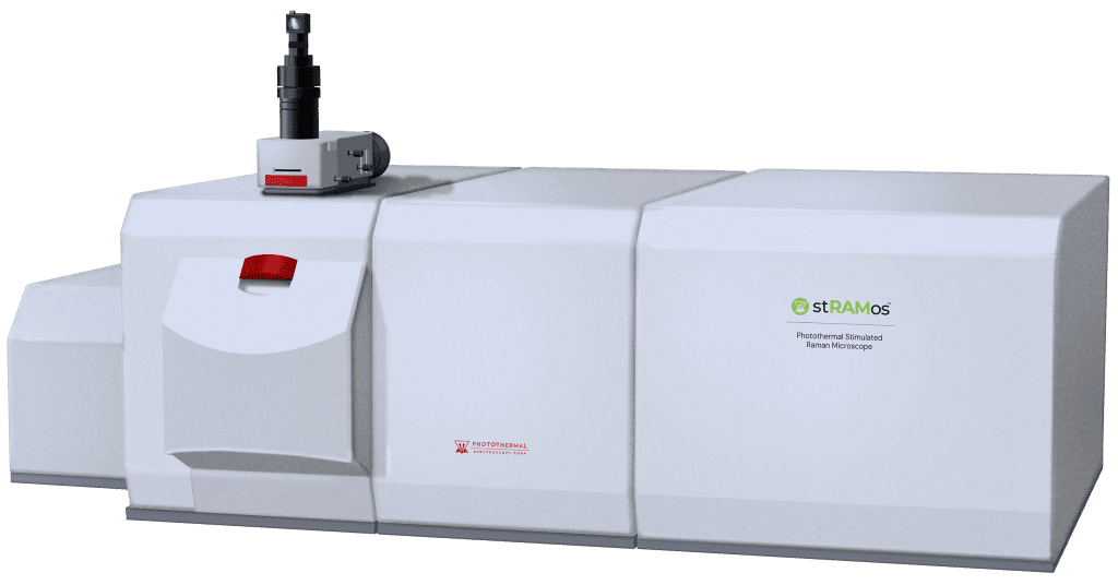
stRAMos™
Sub-300nm Photothermal Stimulated Raman Microscope. Powered by PhotoThermalSRS™
Highest sensitivity measurements - 10x improvement over SRS
≤300nm Raman chemical spatial resolution
High speed chemical imaging for live cells and molecular dynamics
About stRAMos
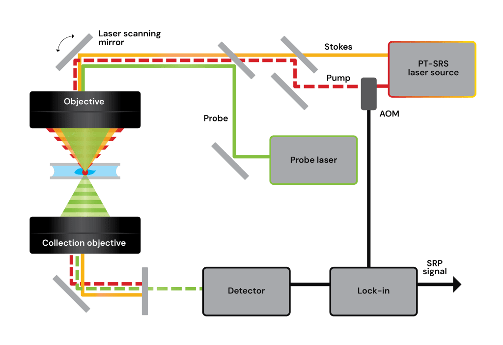
The stRAMos Photothermal Stimulated Raman Microscope uses the PhotoThermalSRS™ (PT-SRS) technique to deliver the combined highest measurement sensitivity and spatial resolution for chemical imaging in a single system with high imaging speeds and advanced microscopy characterization capabilities.
The stRAMos microscope also uniquely combines many optional multimodal capabilities into a single system. stRAMos supports, the combination of PT-SRS with Optical Photothermal Infrared Spectroscopy (O-PTIR), widefield Epi Fluorescence imaging and spontaneous Raman spectroscopy, enabling the most thorough chemical characterization of your sample.
stRAMos with its multimodal capabilities provides the most comprehensive chemical imaging and spectroscopy characterization for a wide range of life science applications including both dry and live cell imaging, biomolecular dynamics of proteins, lipids and other biomolecules.
stRAMos key capabilities
The following videos showcase the stRAMos system on yeast cells prepared in sugar, which increased lipid production. PT-SRS enables high-speed imaging of lipids and proteins in the cells.

Hyperspectral Chemical Imaging in Live Cells
PT-SRS enables full hyperspectral imaging across both the C–H and fingerprint regions, providing rich chemical contrast without labels. By capturing spectra at every pixel, researchers can distinguish subtle but critical molecular variations within live cells.
This capability goes beyond single-wavelength imaging, allowing chemical fingerprints to be mapped across entire populations of cells. The result is a comprehensive view of cellular composition and dynamics — revealing heterogeneity, metabolic states, and molecular organization that would otherwise remain hidden.
Hyperspectral imaging of live yeast cells from 2800cm-1 to 3017cm-1
High Speed, Label Free Imaging for Molecular Dynamics
By enabling imaging at multiple frame rates than conventional techniques, stRAMos makes it possible to study biologically and chemically significant, molecular events. Many biomolecular dynamics—such as lipid transport, protein conformational changes, or membrane reorganization—occur on timescales where conventional imaging methods provide limited insight.
With its unique ability to sensitively capture vibrational signals without labels, stRAMos bridges this gap, revealing dynamic processes that unfold over milliseconds to seconds. This opens new opportunities to monitor molecular reorganization, track metabolic activity, and investigate functional changes in living systems with unmatched clarity.
High speed single wavelength imaging of cells. Imaging of lipids measured at 2850cm-1.
Volumetric chemical sectioning with submicron resolution
stRAMos enables 3D chemical mapping of cells with unprecedented clarity. Unlike conventional volumetric methods, which often compromise resolution, stRAMos delivers true submicron sectional imaging. This allows researchers to resolve fine structural and biochemical details within cells, revealing organization and molecular composition layer by layer. By combining chemical specificity with high-resolution sectioning, stRAMos provides a powerful new way to study cellular architecture and molecular distribution in three dimensions — without labels or staining.
3D Volumetric imaging of lipids and proteins in a yeast cell. Video of biomolecules measured at multiple depths at the fixed wavenumber of 2850cm-1. Image depth varies down to 17µm. Image slightly speeded up.
Multimodal imaging with correlative O-PTIR and Fluorescence Microscopy
The stRAMos system uniquely combines optional multimodal capabilities into one platform. In a combined system, the stRAMos supports PhotoThermalSRS with Optical Photothermal Infrared Spectroscopy (O-PTIR), Widefield Epi Fluorescence microscopy and spontaneous Raman spectroscopy.
Webinar:
PhotoThermalSRS: Faster, more sensitive and robust Stimulated Raman Scattering imaging
More products

Sub-500nm Laser Scanning O-PTIR Microscope for High-Speed Imaging. With co-located Fluorescence and Raman spectroscopy.

The mIRage-LS addresses both life science applications and the broad application set of multiuser facilities where the most advanced vibrational spectroscopy needs are required.

The mIRage platform is modular microscope that delivers high performance submicron IR spectroscopy and imaging with a range of options to address broad application flexibility.
Need more information?
Discover how O-PTIR technology can elevate your research or help solve your toughest challenges. Our team are happy to assist and answer your questions.