
PhotoThermalSRS Technique
PhotoThermalSRS (PT-SRS) represents a breakthrough in label-free microscopy, combining the chemical specificity of Stimulated Raman Scattering with revolutionary photothermal detection. Experience up to 10x higher sensitivity, simplified instrumentation, and unprecedented flexibility for imaging lipids, proteins, and other biomolecules in living systems.
Up to 10× higher than conventional SRS for imaging at real biological concentrations
Robust fiber lasers with easy alignment and long working-distance options
Ultra-low noise detection that avoids the limitations of pulsed lasers
What is PhotoThermalSRS?
Stimulated Raman Scattering (SRS), a form of Coherent Raman Scattering (CRS), is powerful microscopy method producing high speed, high sensitivity, and high spatial resolution label-free chemical imaging. SRS has become an increasingly popular choice in life science research providing chemical imaging of key macromolecules like lipids, proteins, nucleic acids, metabolites, drugs and more in living cells, tissues, organoids and whole organisms—and it continues to gain adoption for rapid, label-free imaging across applications from basic biology to translational studies.
However, the full potential of SRS microscopy has not been realized to date due to many limitations, such as complex instrumentation requiring frequent alignment, insufficient sensitivity, especially at physiologically relevant concentrations and limited sampling flexibility owing to the common need for the use of two immersive, high numerical aperture objectives.
Photothermal Detection Techniques
Photothermal detection of analytical signals, especially with submicron Optical Photothermal Infrared (O-PTIR) spectroscopy has proven its valuable utility with respect to enhanced sensitivity and greatly improved spatial resolution over traditional forms of direct IR spectroscopy, like FTIR and QCL microscopy.
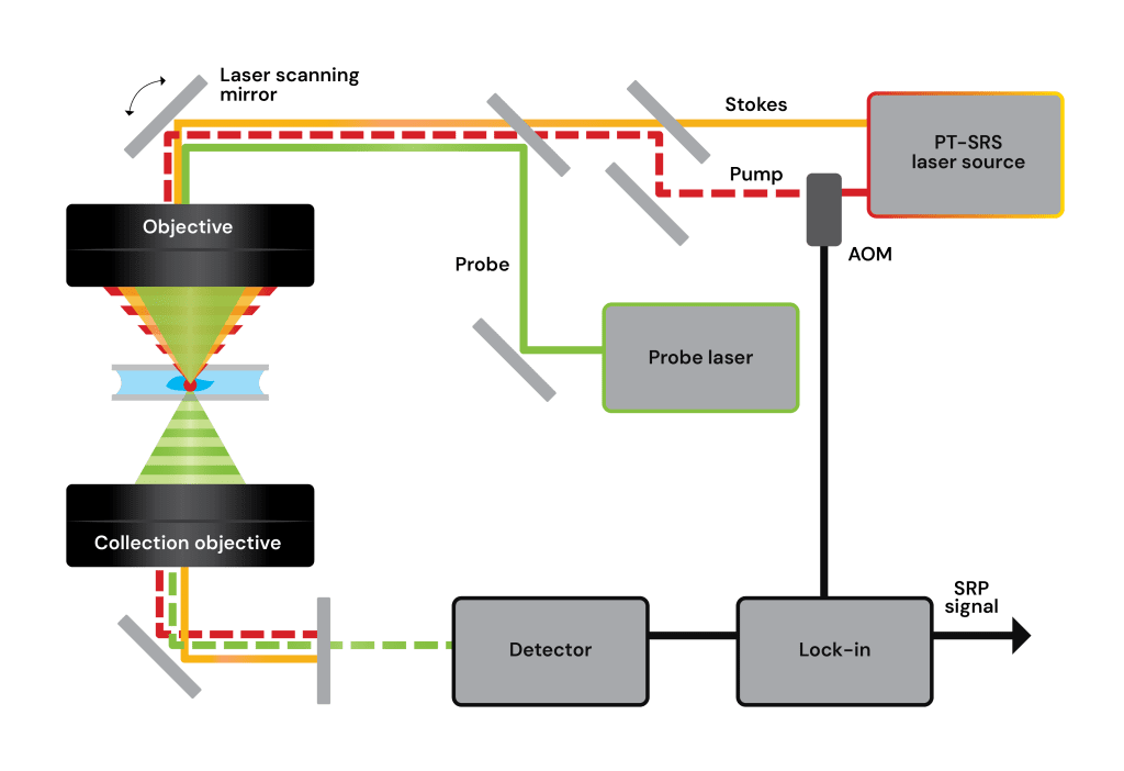
Similarly, photothermal detection is set to revolutionize SRS microscopy with the new breakthrough technique of PhotoThermalSRS (PT-SRS), also known as Stimulated Raman Photothermal, (SRP1). PT-SRS is a new, advanced method of performing Stimulated Raman Scattering (SRS), in which the heat dissipated from the SRS process is detected photothermally with a dedicated ultra-low noise continuous wave (CW) visible (typically 532nm) laser beam, to form the analytical signal, rather than the direct NIR laser light used to generate the SRS effect.
Conventional SRS measurements ignore and do not detect this thermal signal. Instead, SRS is measured by detecting the intensity change of either pump or Stokes laser beams
However, PT-SRS measures intensity changes of the probe which are caused by chemically specific, thermally induced refractive index changes, from non-radiative decay (heat)
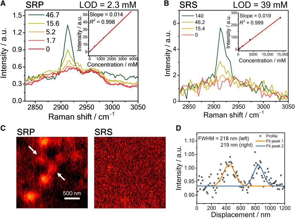
Both the SRS and PT-SRS signals are proportional to band (peak) strength and local concentration, with the PT-SRS signal having added benefit of providing a higher (up to 10x) sensitivity measurement than SRS by thermal accumulation and virtue of the ultra-low noise probe beam, a continuous wave 532nm laser. Because of this, the PT-SRS signal is amplified, and the performance of the SRS laser noise is less of an issue for sensitivity.
Additionally, photothermal detection delivers advantages in experimental flexibility (long working distance condenser for light collection) and use of more robust compact fiber lasers, instead of bulky and complex solid-state lasers.
How PhotoThermalSRS Works
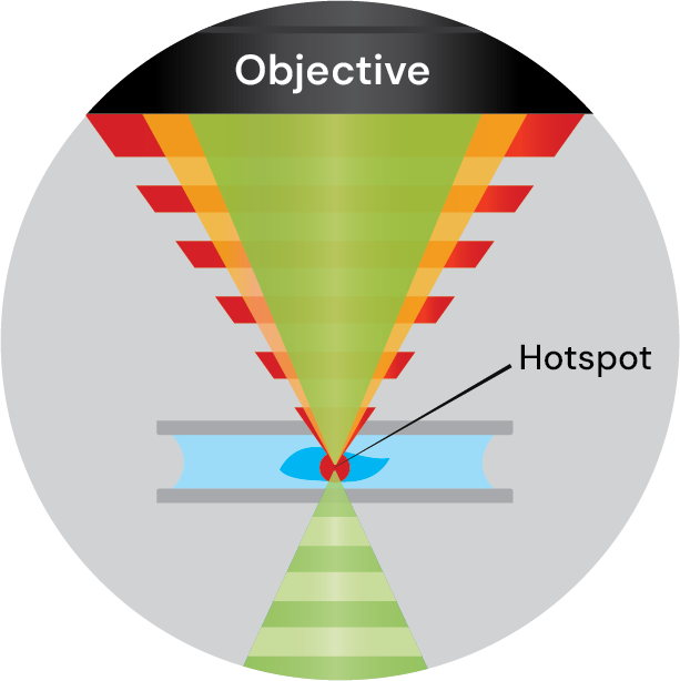
Two tunable NIR picosecond laser beams (Pump and Stokes) are overlapped (in space and time) and directed at the sample.
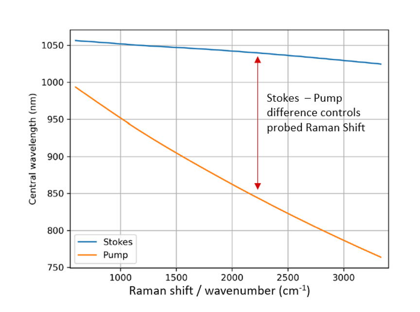
The wavelengths of the Stokes and Pump are tuned such that their optical frequency difference is matched to the vibrational frequency of the target Raman transition (Raman shift).
Changing the wavelength of SRS lasers
When the wavelength gap (optical frequency difference) between the 2 beams exactly matches a molecular bond vibration frequency in the sample, the intensity of the pump beam will decrease, known as stimulated Raman loss (SRL) and the intensity of the Stokes beam will increase, known as stimulated Raman gain (SRG) via the stimulated Raman process.
Image: Changing the wavelengths of SRS lasers (Stokes and Pump) to match target Raman shift.
Cheng, Ji-Xin, et al. “Advanced vibrational microscope for life science.” Nature Methods 22.5 (2025): 912-927
PT-SRS Signal
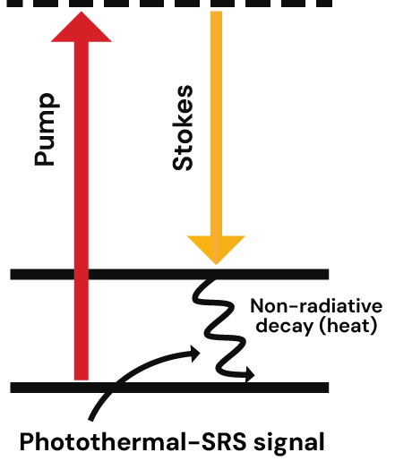
After driving the chemical bond Raman vibration, the vibrational energy is relaxed through non-radiative decay and deposited into the sample as heat, creating changes in thermal expansion and index of refraction. Thermal accumulation is the key to boosting the sensitivity of PT-SRS. Unlike the instantaneous SRS signal (picosecond level) with a small change over the pump/Stokes beams, PT-SRS signal is temporally accumulative since the heat dissipation for molecules in biological samples does not happen within hundreds of nanoseconds or a few microseconds. Thus, the PT-SRS signal can be amplified with the accumulative heat by SRS process.
Since the NIR pump beam is (externally) modulated (typically to a few to several hundred kHz), the changes in expansion and index of refraction are also modulated at the same frequency. These are then transferred to modulations in the transmitted visible probe laser, with the analytical signals being extracted via a lock-in amplifier, resulting in a highly sensitive photothermal detection mechanism.

Impact of laser noise on sensitivity
The sensitivity of SRS is fundamentally limited by the relative intensity noise (RIN) of pulsed NIR lasers, since the signal is measured as a small modulation of the transmitted pump or Stokes beam. Ideally, the chemical sensitivity for SRS should be shot noise limited. In practice, the complex noise sources inherent to pulsed lasers limits the sensitivity performance of SRS.
In conventional SRS microscopy, to achieve a sensitivity close to shot noise limit, femtosecond, or picosecond mode-locked, complex solid-state lasers are typically used to generate the synchronized pump and Stokes beams, with one beam modulated for lock-in detection.
An alternative to these complex solid state lasers is the fiber laser. Picosecond NIR fiber lasers have recently become commercially available and offer considerable advantages, being simple, robust, compact, and very importantly for SRS, having extremely fast wavelength tuning (25-100ms)
Image: Stimulated Raman Photothermal microscopy breaks the fundamental shot-noise barrier of SRS, delivering > 100× higher chemical SNR and low-mM / µM detection limits with simpler, fiber-laser hardware and relaxed optical constraints.
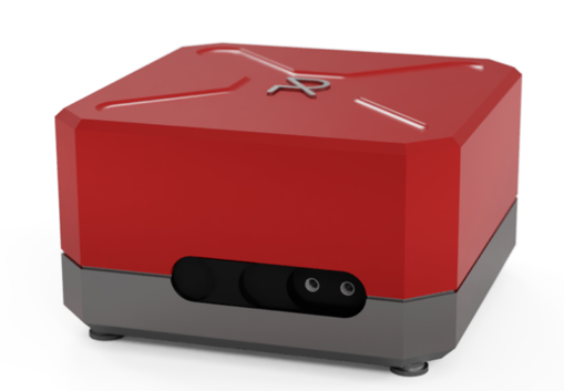
Compact, high speed, high spectral resolution laser.
Whilst these advantages are useful, fiber lasers have not gained widespread SRS usage as these are often not shot noise limited, resulting in poorer sensitivity when used with traditional SRS setups. In contrast, the key enabler for fiber lasers for SRS, comes through Photothermal detection of the SRS signal – PhotothermalSRS. PT-SRS uses fiber lasers for the pump and Stokes excitation. Raman-induced absorption is read out via a separate ultra-low noise continuous-wave (CW) probe laser.
Because the photothermal signal is encoded in refractive index or scattering changes sensed by the CW probe, the sensitivity (1) can be improved by thermal accumulation which cannot be achieved by conventional SRS, and (2) is not limited by noise in the pulsed pump/Stokes lasers but instead by the ultra-low noise of the CW probe. As a result, PT-SRS can achieve high sensitivity without being impacted by the high RIN typical fiber laser sources, whilst being able to deliver on all of the benefits of fiber lasers.
Publication:
Stimulated Raman photothermal microscopy toward ultrasensitive chemical imaging
Zhu et al., Sci. Adv.
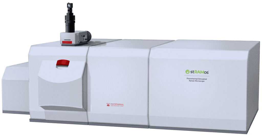
Sub-300nm Photothermal Stimulated Raman Microscope. Powered by PhotoThermalSRS™
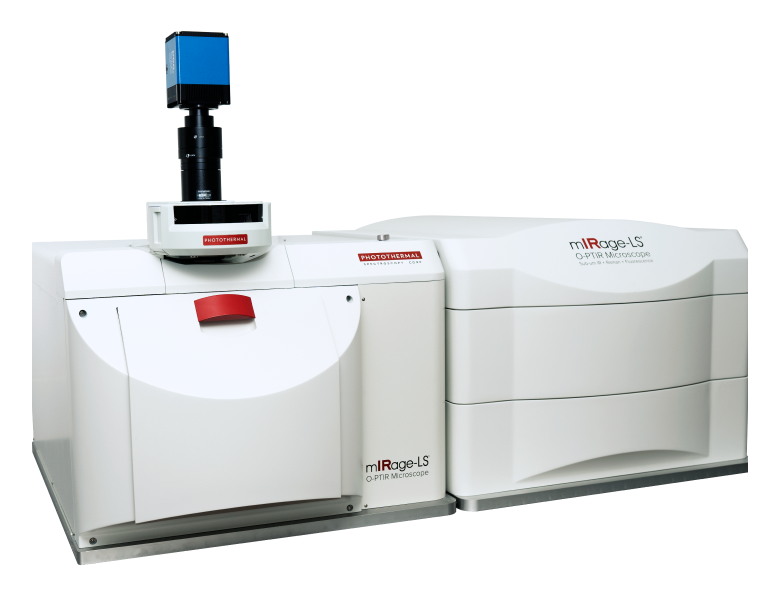
The mIRage-LS addresses both life science applications and the broad application set of multiuser facilities where the most advanced vibrational spectroscopy needs are required.

The mIRage platform is modular microscope that delivers high performance submicron IR spectroscopy and imaging with a range of options to address broad application flexibility.
Need more information?
Discover how O-PTIR technology can elevate your research or help solve your toughest challenges. Our team are happy to assist and answer your questions.