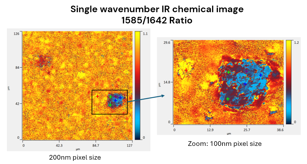
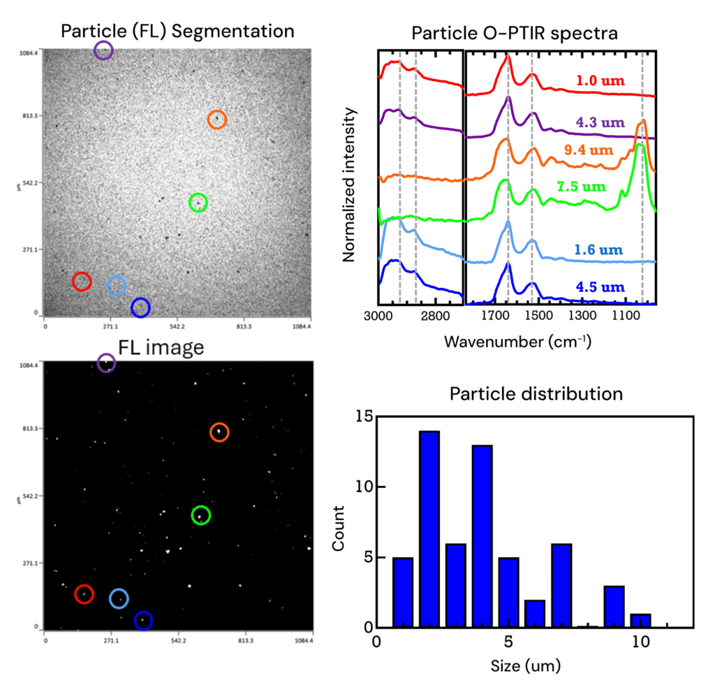
Submicron IR for pharmaceuticals

Detection and chemical ID of smaller sub-visible, even submicron particulates in pharmaceutical formulations is becoming increasingly important. Submicron, multi-modal O-PTIR addresses this problem with its unique approach to spectroscopy and microscopy of defects and particulates.

Why O-PTIR for pharmaceutical research?

Particulate matter in sterile pharmaceutical formulations presents a serious health challenge with potentially severe consequences for patients. Additionally, protein aggregates and proteins adsorbed to foreign microparticles in therapeutic protein products increase immunogenicity, diminishing therapeutic response. Consequently, regulatory authorities are requesting that the characterization and visualization of submicron particles be incorporated into the particle control strategies. 

Datasheet:

mIRage®-Raman microscope

Automated Raman today. Multimodal Spectroscopy tomorrow.

Particulate matter in protein therapeutics formulations

Integrating submicron chemical and morphological analysis into routine particle control strategies is now a reality with mIRage-LS, submicron multimodal IR microscope. Routine analysis of submicron chemical and morphological analysis of particulate matter and particles enables Critical Quality Attributes (CQA) to be established that contribute to quality, safety and efficacy of infusion and injection formulations. 

Moreover, submicron (O-PTIR) spectra collected on highly fluorescent biologic matrices, traditionally considered challenging to impossible with tools like Raman microscopy, are now completely unaffected, enabling robust submicron chemical characterization and particle visualization of aggregates and particulate matter.

Here we describe how multi-modal O-PTIR has been employed to investigate Particulate Matter in Protein Therapeutics Formulations

Composite particles: single-frequency chemical imaging

  • Chemical image reveals ~5 µm FFA particle (with protein) and ~20 µm proteinaceous aggregate (with FFA inclusions weighted to the left-hand side of the particle)
  • Sub-micron IR chemical imaging has shown intraparticle heterogeneity
Composite Particles: Single-Frequency Chemical Imaging
Fluorescence imaging & IR spectroscopy

Fluorescence imaging & IR spectroscopy

Whilst filtered particulate matter in the brightfield image has limited contrast, the biological nature of these protein aggregates provides for strong autofluorescence imaging, significantly increasing their contrast, allowing for more accurate particle sizing and statistics as well as providing for more accurate locating for subsequent IR spectroscopy.

The sub-micron IR (O-PTIR) spectra are collected from particles as small as ~1 µm in size with excellent spectral sensitivity with measurements times less the 5 sec per spectrum.

Clear secondary structural differences are evident based on the differences in shape of the Amide I band (~1660 cm-1) with these differences being most prominent for the larger particles which also spectral differences at ~1000 cm-1.

Protein specific IR chemical imaging & IR spectroscopy

Ratioed single frequency ratio images (1641/1587 cm-1) highlight only the proteinaceous particles providing for a more selective, targeted for protein chemical image. Full range IR (O-PTIR) spectra of these protein particles shows differences in protein secondary structure (beta sheet vs alpha helical) with differences in silicon oil content also being observed

Protein specific IR chemical imaging & IR spectroscopy

White paper:

O-PTIR of biopharmaceutical particulates

Webinar:

Submicron IR+Raman analysis of biopharmaceuticals

Datasheet:

mIRage: Multimodal submicron O-PTIR microscope

Technical article:

Advancing biopharmaceutical analysis with simultaneous submicron IR and Raman spectroscopy

Webinars

Need more information?

Discover how O-PTIR technology can elevate your research or help solve your toughest challenges. Our team are happy to assist and answer your questions.

Fill out the form to watch the webinar

No video available.