30x faster O-PTIR: Hyperspectral imaging in minutes,
single wavelength imaging in seconds
Sub-500nm IR chemical spatial resolution
Multi-modal correlative fluorescence imaging and Raman spectroscopy
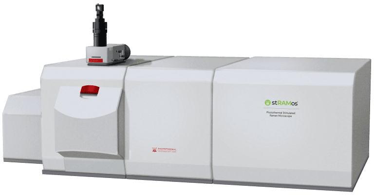
About mIRage-HSi
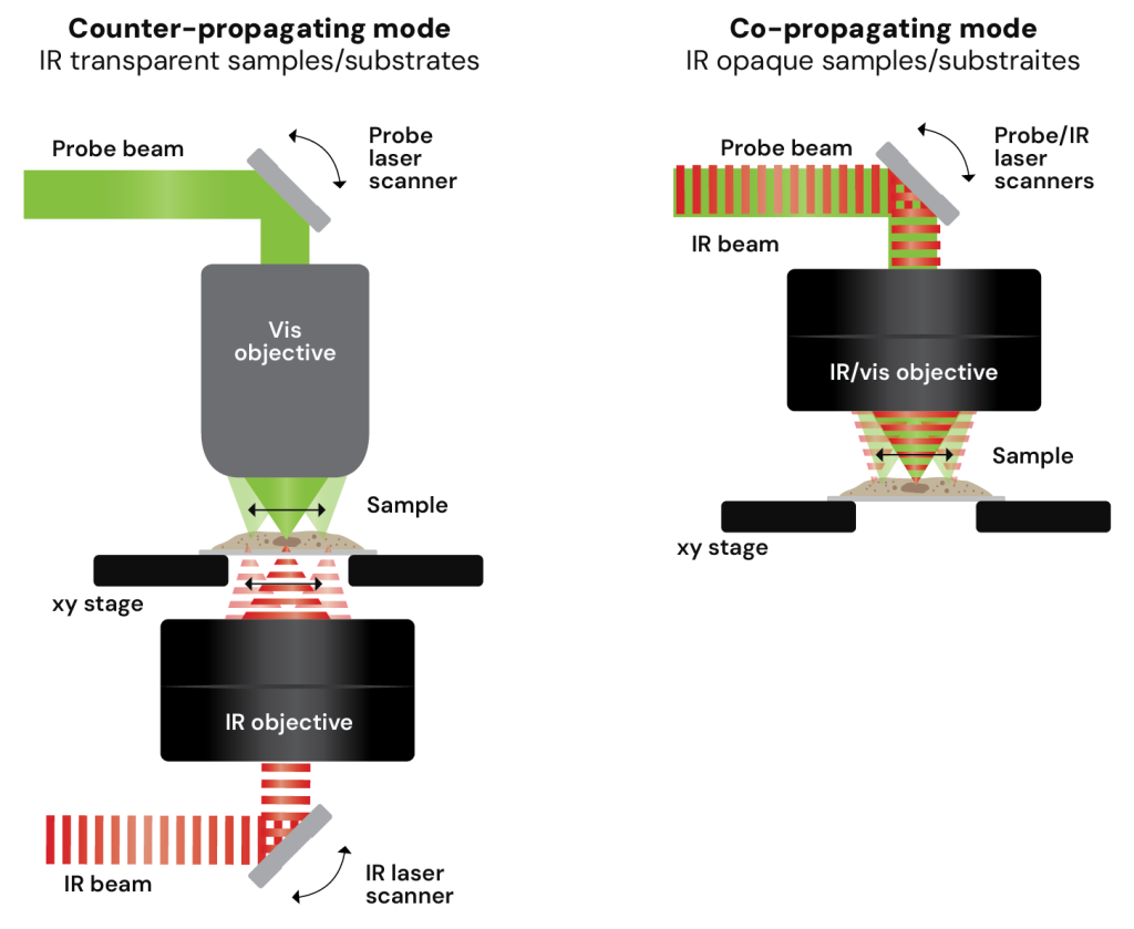
Next-level performance: Laser scanning O-PTIR
The new mIRage-HSi is the next evolution of Optical Photothermal Infrared (O-PTIR) spectroscopy and imaging, delivering the highest performance sub-500nm IR imaging and FTIR transmission/ATR-like spectroscopy but now with breakthrough speed reducing hyperspectral imaging time from hours to minutes and single frequency images from minutes to seconds.
This dramatic increase in imaging speed extends O-PTIR technology to dynamic studies, higher speed contaminant detection, and automated submicron particle identification.
Whether for microplastics, pharma, life science, failure analysis, or materials science, mIRage-HSi delivers faster, more automated, and more productive O-PTIR workflows.
Learn more about how the mIRage-HSi can help your research reach new levels.
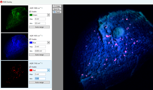
PTIR RGB overlay provides an automated measurement method for user selectable wavenumbers with resulting RGB overlay
The RGB overlay example shows 3 distinct wavenumbers representing:
1080cm-1 – Nucleic acid
1655cm-1 – Proteins
1738cm-1 – Lipids
Each wavenumber took 16 seconds.
Preview mode: real-time chemical imaging for ROI significant EoU and productivity improvement
Preview mode is a lower resolution, high speed chemical imaging preview of a selected area.
It provides a significant enhancement in measurement productivity for locating the feature of interest.
Move, Locate, Image
Quickly identify the relevant ROI for defects, contaminants, microplastics over an area greater than the objective FOV.
Application overview
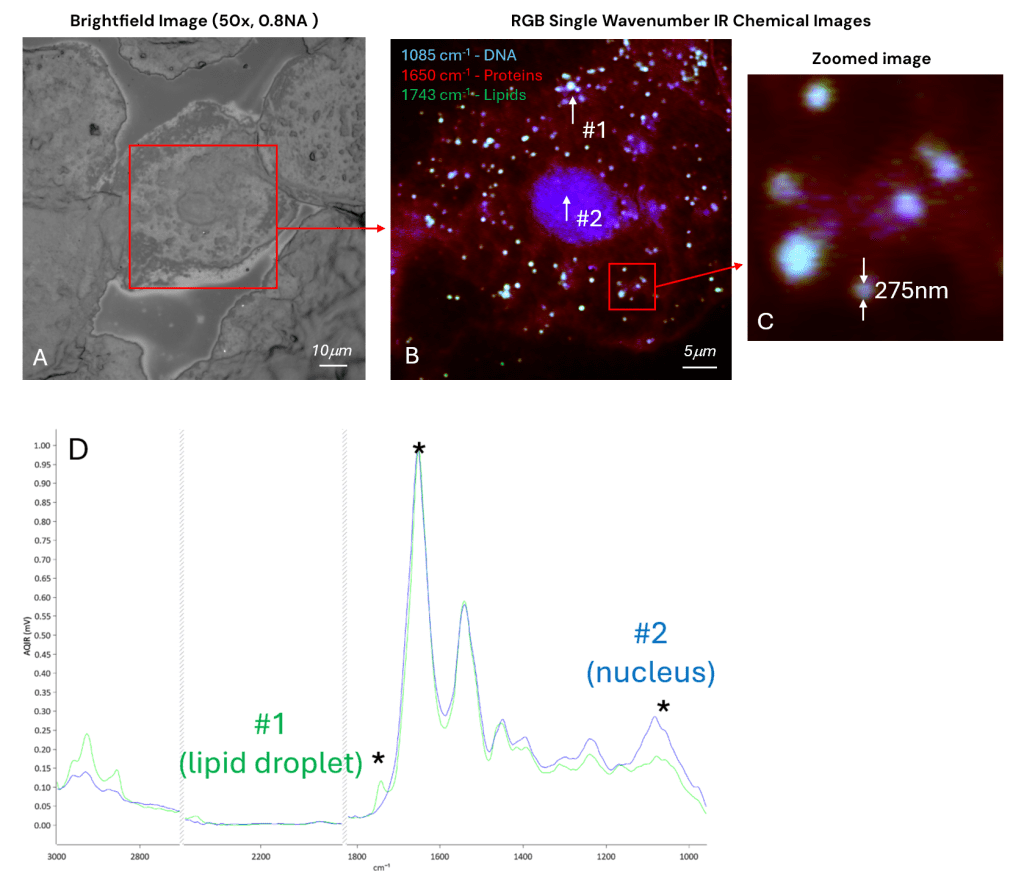
13 sec per image measurement time
High-speed chemical imaging of cheek cells
A: Brightfield image using a 50x, 0.8NA objective B: Single wavenumber IR chemical imaging RGB overlay, 1085cm-1 DNA (blue), 1650 cm-1 protein (red) and 1743 cm-1 lipid (green) C: Zoomed in image of B, showing detection of lipid droplets as small as 275nm D: O-PTIR Spectra (normalised to Amide I) from highlighted locations, spot #1 (lipid droplet) and spot #2 (nucleus). IR single wavenumber images (as in B) are market by an asterisks. Spectra are collected from a spot size of ~500nm.
Collection conditions: 50×50µm measurement FOV, 100nm pixel step size (250,000 pixels), 13 sec per image measurement time, Counter-prop mode. Spectral acquisition total time 8 sec 50x, 0.8NA glass objective, tri-range IR QCL, 532nm probe.
Sample courtesy of Dr. Mark Hackett, Curtin University, Australia.
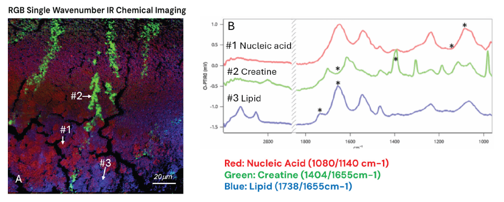
17 sec per image measurement time
High speed multi-spectral IR imaging of mouse brain tissue
A: Automated multiple (5) single wavenumber IR chemical imaging RGB overlay – Green (Creatine) 1404/1655cm-1), Red (Nucleic Acid) 1080/1140 cm-1 and Blue (lipid) 1738/1655cm-1 B: Representative spectra from the highlighted locations, highlighting biochemistry from areas of chemical contrast as shown in A. IR image (A) wavenumber positions are marked by an asterisks. Spectra are collected from a spot size of ~500nm.
Collection conditions: 150×150µm measurement FOV, 250nm pixel step size (360,000 pixels), 17 sec per image measurement time, counter-prop mode with 50x, 0.8NA glass objective, dual-range IR QCL, 532nm probe.
The mIRage-LS addresses both life science applications and the broad application set of multiuser facilities where the most advanced vibrational spectroscopy needs are required.
The mIRage platform is modular microscope that delivers high performance submicron IR spectroscopy and imaging with a range of options to address broad application flexibility.
Discover how O-PTIR technology can elevate your research or help solve your toughest challenges. Our team are happy to assist and answer your questions.