
Publication link
“Widefield Super-Resolution Infrared Spectroscopy and Imaging of Autofluorescent Biological Materials and Photosynthetic Microorganisms Using Fluorescence Detected Photothermal Infrared (FL-PTIR),”
The authors present a novel approach for high-speed, super-resolution infrared (IR) spectroscopy and chemical imaging of autofluorescent biological materials using fluorescence detected photothermal infrared (FL-PTIR). This method leverages temperature-dependent modulations in autofluorescent emissions to achieve high spatial resolution and fast data acquisition. The study focuses on materials like collagen, plant tissues, diatoms, and green microalgae, highlighting the potential for dynamic, label-free imaging of live cells and tissues in water. This work showcases the ability of FL-PTIR to provide detailed chemical mapping with sub-500 nm resolution, significantly improving upon conventional techniques.
Specific data supporting the FL-PTIR approach include the examination of collagen, which exhibits strong autofluorescence due to tyrosine residues and cross-links. The study provided detailed IR chemical images and spectra of collagen fibrils under different polarizations, revealing molecular orientation effects. Another example is the application on photosynthetic organisms, where the intrinsic autofluorescence of chlorophyll allowed for high-resolution spectroscopic measurements of plant tissues and photosynthetic microorganisms. This was demonstrated through composite IR absorption images and spectra of plant tissue, showcasing the distribution of different chemical constituents.
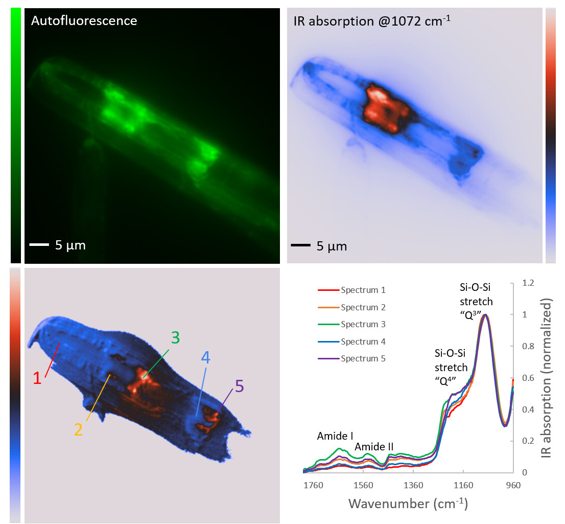
The research further explored the capabilities of FL-PTIR with diatoms, (refer to figure) which are photosynthetic microalgae with silica-based cell walls. By analyzing the autofluorescence from chlorophyll and silica, the study mapped chemical variations in diatom frustules at sub-micrometer scales. The hyperspectral data collected provided insights into the silicon-oxygen bonding and protein distribution within the diatom structures. This level of detailed chemical imaging highlights the effectiveness of FL-PTIR in studying complex biological samples.
In conclusion, the authors note that “FL-PTIR is a powerful tool for rapid, high-resolution chemical imaging of autofluorescent biological materials. This technique offers significant advantages over traditional methods by providing faster data acquisition and higher spatial resolution without the need for external labels”.
Further the ability to perform dynamic measurements on live samples, such as green microalgae in water, opens new possibilities for studying biochemical processes in real-time. The authors suggest that FL-PTIR could be widely applicable in various fields, including biomedical research, environmental monitoring, and materials science.
Dr. A. Banas, Dr. K. Banas
Prof. R. Chandramohanadas
Singapore Synchrotron Light Source (SSLS)
NUS, Singapore
Communication Chemistry, 2021.
DOWNLOAD PDF
