
“This study implements O-PTIR spectroscopy for a new application, providing label-free non-destructive chemical imaging of functional groups throughout biofilm depth. Our work showed that biofilms have protein synthesis gradients using this chemical imaging capability. Using the O-PTIR platform to investigate antimicrobial treatments gives detailed information about the effect a treatment has on biofilm protein metabolism, demonstrating the impact antibiotic treatment has on this metabolism as well as the protective properties biofilms confer against treatment.”
FULL PUBLICATION >
Reporting in Communications Biology, researchers at the University of Liverpool have introduced a new implementation of optical photothermal infrared (O-PTIR) spectroscopy for the spatially resolved, label-free chemical imaging of bacterial biofilm metabolism — and applied it to understand how antibiotic treatment reshapes metabolic activity inside intact Salmonella Typhimurium biofilms.
Bacterial biofilms are a critical healthcare problem: their layered architecture generates metabolic gradients that promote antibiotic tolerance and antimicrobial resistance (AMR). Metabolically dormant cells at the biofilm core downregulate protein synthesis pathways — key antibiotic targets — while more active cells occupy the periphery. Yet most analytical methods either rely on bulk measurements that lose spatial context, or on fluorescent labels that can perturb the biology. Obtaining chemically specific, spatially resolved information about metabolism at depth within an intact biofilm — and observing how it changes under antibiotic challenge — has remained a significant technical gap.
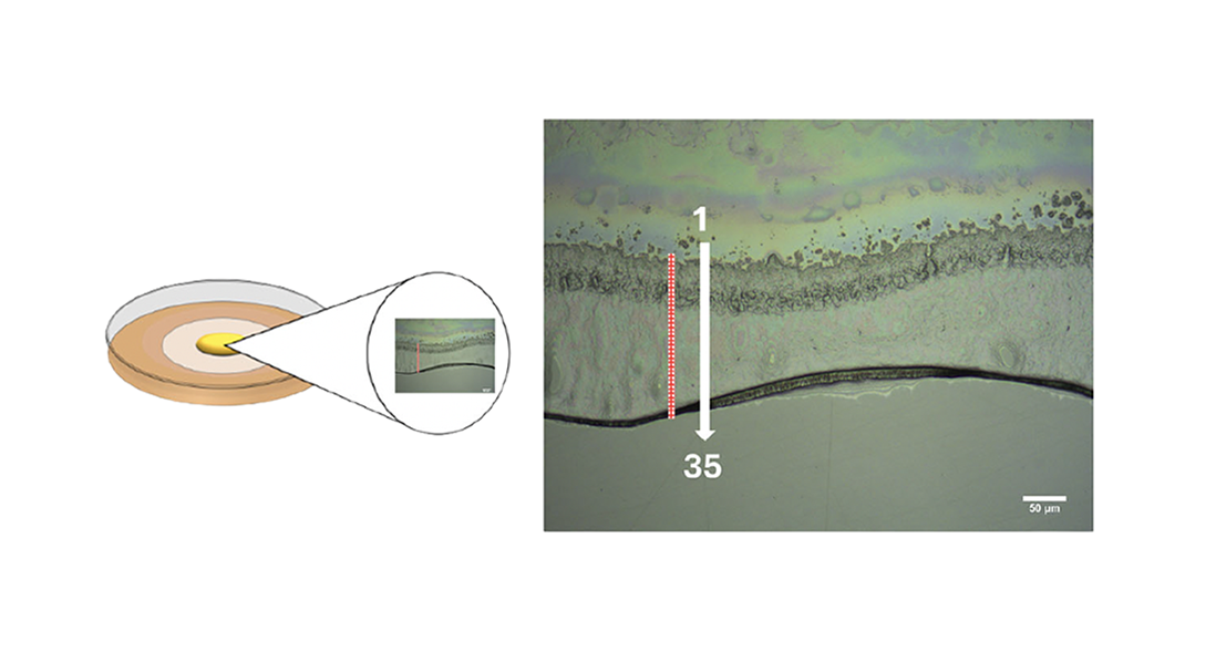
The team addressed this by pairing O-PTIR spectroscopy with ¹³C stable isotope probing (SIP) and an optimised cryosectioning protocol. Biofilms of S. Typhimurium 4/74 were grown on polycarbonate membranes and then transferred to minimal medium containing uniformly labelled [U-¹³C₆]-D-glucose as the sole carbon source, with or without antibiotic supplementation. After 24 hours, biofilms were cryosectioned to 5 µm and imaged on CaF₂ slides. O-PTIR single-frequency imaging at 1655 cm⁻¹ (¹²C amide I) and 1616 cm⁻¹ (¹³C amide I) produced ratiometric chemical maps of protein synthesis activity across the biofilm cross-section at ~500 nm spatial resolution.
Because O-PTIR uses a visible-light probe (532 nm) to detect mid-IR photothermal signals, it overcomes the diffraction limit of conventional FTIR microspectroscopy — enabling cell-scale chemical imaging that conventional IR cannot achieve. Point spectra collected at 5 µm intervals through the biofilm depth and analysed by principal component analysis (PCA) complemented the imaging data.
In untreated biofilms, ratiometric O-PTIR imaging revealed a core of cells with minimal ¹³C incorporation surrounded by a metabolically active periphery — consistent with oxygen-driven stratification. PCA of depth-resolved spectra showed that the amide I band, accounting for 82.31% of total explained variance (PC1), was the dominant discriminator between high- and low-metabolism regions, with additional contributions from amide II, lipid/amino acid, DNA, and carbohydrate peaks.
When biofilms were treated with gentamicin — to which the engineered strain was non-susceptible — the metabolic gradient was preserved, with three PCA clusters (high, intermediate, and low ¹³C incorporation) explaining 88.17% of variance. Kanamycin treatment, by contrast, markedly disrupted ¹³C incorporation across the biofilm: no incorporation was detected at the biofilm base where antibiotic concentration was highest, and only limited incorporation was observed at the uppermost layer. PCA revealed two clearly separated clusters (75.9% TEV), and the PC1 loadings also highlighted a symmetric PO₂⁻ stretching peak from DNA — suggesting broader biochemical changes beyond metabolic suppression in antibiotic-stressed cells.
This study demonstrates that the combination of O-PTIR imaging with ¹³C SIP and cryosectioning delivers a uniquely capable platform for biofilm metabolic analysis. It is the first application of ¹³C SIP with spatially resolved O-PTIR imaging in a biofilm context, and the first to visualise carbon metabolic gradients at ~500 nm resolution using ratiometric O-PTIR imaging. The approach is label-free, non-destructive, and chemically specific — enabling simultaneous visualisation of metabolic heterogeneity across the full depth of a biofilm cross-section while preserving native architecture.
The results validate O-PTIR as a tool for assessing antibiotic efficacy against biofilms with spatial resolution, and point toward future applications in mixed-species biofilms, shared-resistance mechanisms, flow-cell biofilm models, and integration with complementary mass spectrometry imaging modalities.
Authors:
Daniel Smaje, Xiaojun Zhu, Jay C. D. Hinton, Rasmita Raval, Royston Goodacre, Howbeer Muhamadali
