
Microplastics
Enabling submicron microplastics and nanoplastics analysis for human health research
The unique technology of sub-500nm IR and Raman Optical Vibrational Photothermal Microscopy and spectroscopy delivers automated chemical identification of nanoplastics with the highest chemical sensitivity and spatial resolution addressing critical measurement requirements of academic researchers and government directed funding.
Multi-modal Optical Photothermal IR Spectroscopy (O-PTIR) has demonstrated unrivalled resolution, performance and flexibility for microplastics measurements relating to human health for measurement in cells and tissues with detection sensitivity to ~200nm in size.
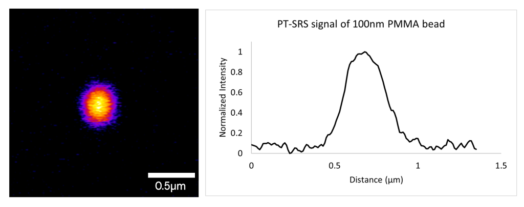
The sensitivity and spatial resolution of Photothermal Stimulated Raman Scattering (PT-SRS) excels at live cell imaging and provides measurements of microplastics with up to 100nm detection capability.
Now both laser-scanning based O-PTIR and PT-SRS are available on one combined platform the stRAMos to address the broadest range applications in human health and microplastics.
Why new measurement approaches are needed
Understanding the impact of microplastics on human health requires reliable detection and chemical identification at the smallest scales. However, most conventional techniques face critical limitations:
- Raman microscopy is limited by fluorescence interference, low sensitivity and is prone to misidentifying fats/lipids/stearates with PE
- Traditional Direct IR (FTIR/LDIR/QCL) microscopy is constrained by diffraction limits (~10 µm), missing smaller particles and intracellular biochemical detail
- SRS microscopy is hindered by complex instrumentation, limited chemical specificity and spectral range, and restricted sampling options.
- Pyrolysis GC-MS is destructive, lacks spatial information and is prone to misidentification of some polymers like PE and PVC with biological materials
Thus, submicron and nanoplastics, which are the most biologically relevant size fraction, remain difficult to measure and identify.
These gaps limit the ability to study microplastics in biological systems, where particles are often small, heterogeneous, and embedded in complex matrices.
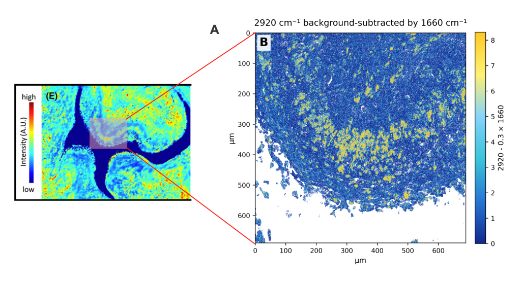
O-PTIR sub-1 µm resolution tissue imaging (right) vs FTIR imaging (resolution 10µm) (left)
How the mIRage multimodal O-PTIR products overcomes these challenges
The mIRage™ O-PTIR based spectroscopy and microscopy products uniquely enable chemical identification and imaging and sizing of micro- and nanoplastics at sub 500nm scales while providing biochemical information on the impact on cells and tissues of the presence of these particles.
- Submicron resolution detection (~200 nm): Detect and identify particles and biochemical cellular responses below the limits of traditional IR & Raman spectroscopy
- O-PTIR provides optical measurements (non-contact, no ATR) with FTIR transmission/ATR-like spectra, collected in reflection mode.
- Direct Analysis in MPs/NPs in cells and tissues with simultaneous biochemical insights
- featurefindIRTM workflow that addresses automation and ease of use for particle selection and identification
- Unique multi-modal capabilities leveraging co-located widefield and/or confocal fluorescence microscopy to align with existing life science workflows and for providing targeted regions of interest for O-PTIR chemical analysis.
This combination allows researchers to study microplastics in biological environments with greater confidence, particularly at the size scales most relevant to human health.
Read below to learn more about why the National Institute of Standards and the US FDA selected the mIRage product for microplastics measurements.
mIRage with O-PTIR continues to gain widespread adoption for microplastics research in academia, industry, and government, with recent installations at NIST, FDA and leading microplastics research institutions.
The new mIRage HSi, provides higher speed measurements.
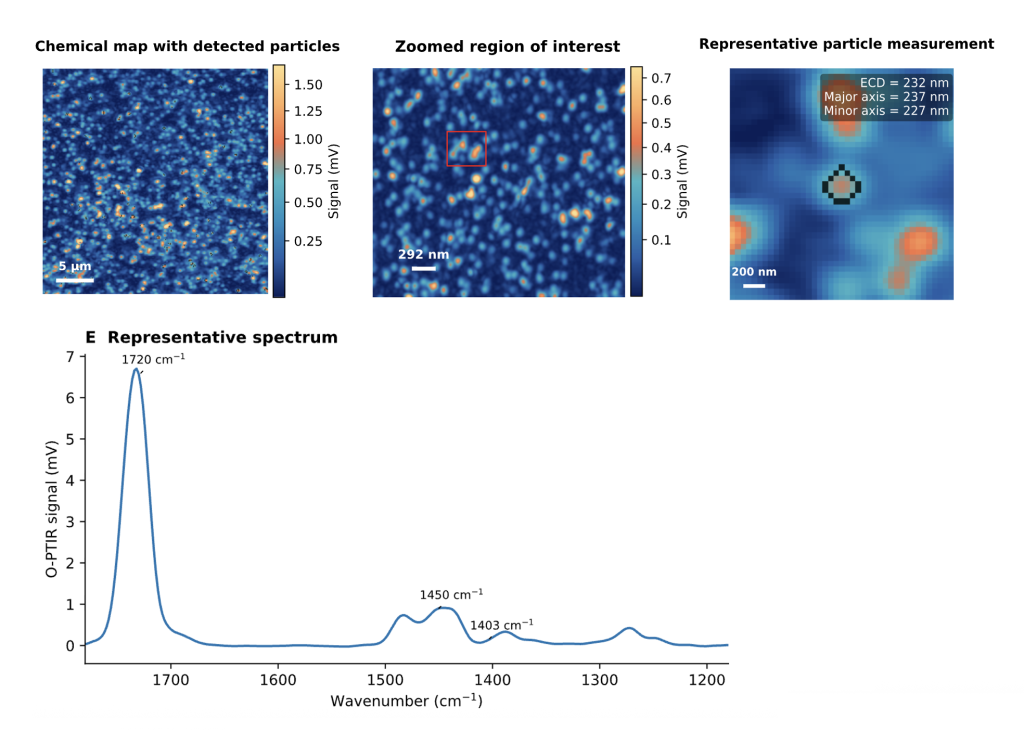
250nm PMMA polymer beads on. 200nm pore size Anodisc (Alumina) filters
O-PTIR performance for nanoplastics imaging in tissues
<500 nm
Infrared spatial resolution
Glass slides
compatible reflection geometry
IR+Raman+FL
co-located multimodal confirmation
O-PTIR ~200nm detection sensitivity
PT-SRS ~100nm detection sensitivity
Identifying microplastics in tissues & cells
What is it? Separates out the small nanoplastics from the tissue.
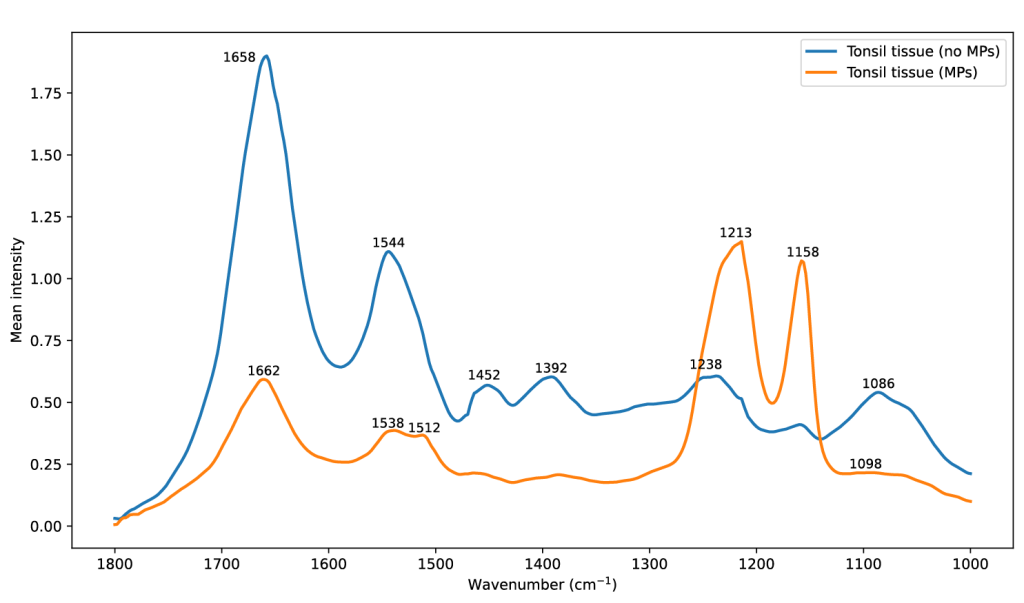
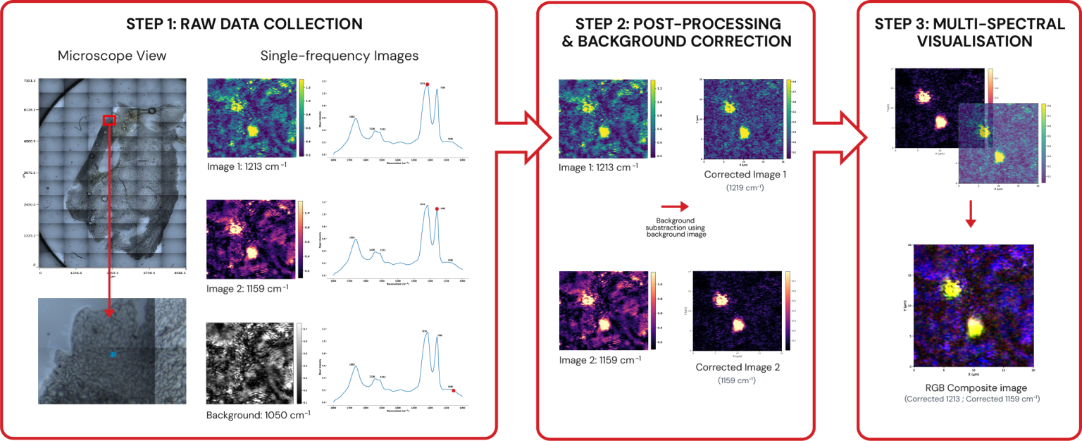
Comparison of mean spectra from tissue regions containing nanoplastics (orange) versus adjacent nanoplastic-free (blue) regions. The nanoplastic-containing regions (orange) exhibit distinct peaks at ~1213 cm⁻¹ and ~1159 cm⁻¹, corresponding to asymmetric and symmetric C–F stretching, respectively. These bands are absent from surrounding tissue and do not overlap with amide I/II or lipid carbonyl features, thereby minimising common sources of false-positive interference
Where is it? High speed single/multi wavelength imaging.
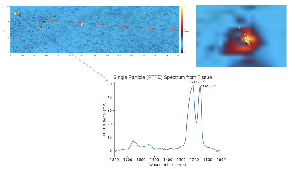
Detection and chemical identification of a ~1.2 µm PTFE microplastic particle in intact formalin-fixed tissue using O-PTIR. This demonstrates that discrete polymer regions as small as ~1 µm can be resolved in situ without extraction or disruption of tissue architecture. Single- (or multi-) frequency chemical imaging (top left) highlights sparse high-intensity particles, while a zoomed region of interest (top right) confirms particle morphology and size. The corresponding single-particle spectrum (bottom), acquired from one of the larger particles, exhibits characteristic PTFE bands at ~1213 and ~1170 cm⁻¹, enabling confident chemical identification.
Nano/microplastic cell interactions
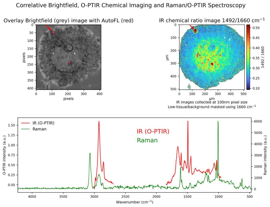
O-PTIR imaging reveals the spatial localization of polystyrene (PS) microbeads within cells using IR chemical contrast, while simultaneous Raman spectroscopy provides complementary molecular confirmation.
Fluorescence signal, from fluorescently labelled PS beads, identifies bead location, and the IR ratio image enhances contrast relative to the cellular background, enabling confident correlation between particle position and biochemical environment.
Sample Courtesy of Prof Gary Hardiman, Queens University Belfast
100nm detection sensitivity
Photothermal SRS complements O-PTIR by extending particle detection sensitivity down to 100 nm, enabling broader micro- and nanoplastics analysis.
“The integration of high-resolution O-PTIR chemical characterization with preserved spatial information provides a promising avenue to elucidate the role of microplastic accumulation in human disease and supports investigation into potential mechanistic links between microplastic exposure and inflammatory processes.”
Publication: Method for label-free & non-destructive detection of microplastics in human formalin-fixed paraffin-embedded tissue sections.
Author: Elizabeth S. Gruber et al.
O-PTIR microplastic tissue workflow: Teflon detection
FEATURED CASE STUDY
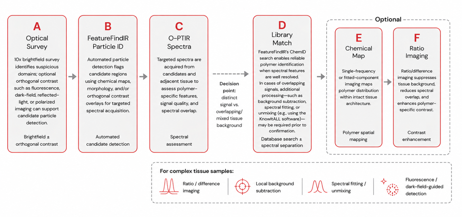
Workflow for in situ polymer identification in complex tissue using O-PTIR and FeatureFindIR
A structured workflow for detecting and identifying polymer particles directly within intact tissue using brightfield screening, supported by optional orthogonal contrast imaging (e.g., fluorescence, dark-field, or polarized light) to aid candidate detection. Automated identification (FeatureFindIR) guides targeted O-PTIR spectral acquisition, followed by ChemID-based library matching for polymer confirmation. Optional chemical mapping and ratio imaging enhance contrast and spatial interpretation. The workflow supports analysis in complex biological environments without extraction, preserving tissue architecture while addressing spectral overlap through background subtraction, spectral fitting, or unmixing.
Learn more about featurefindIR for automated microplastics measurement and chemID
“Our findings demonstrate that sub-500nm O-PTIR is an efficient, non-destructive technique for label-free microplastic studies in biological samples, enabling the identification, localization and characterization of heterogeneous microplastics, while simultaneously revealing their impact on cellular function and composition.”
Publication: Label-free non-destructive spectroscopic detection of mixed microplastic uptake and differential effects on intestinal epithelial cells.
Author: Charlotte E. Sofield et al.
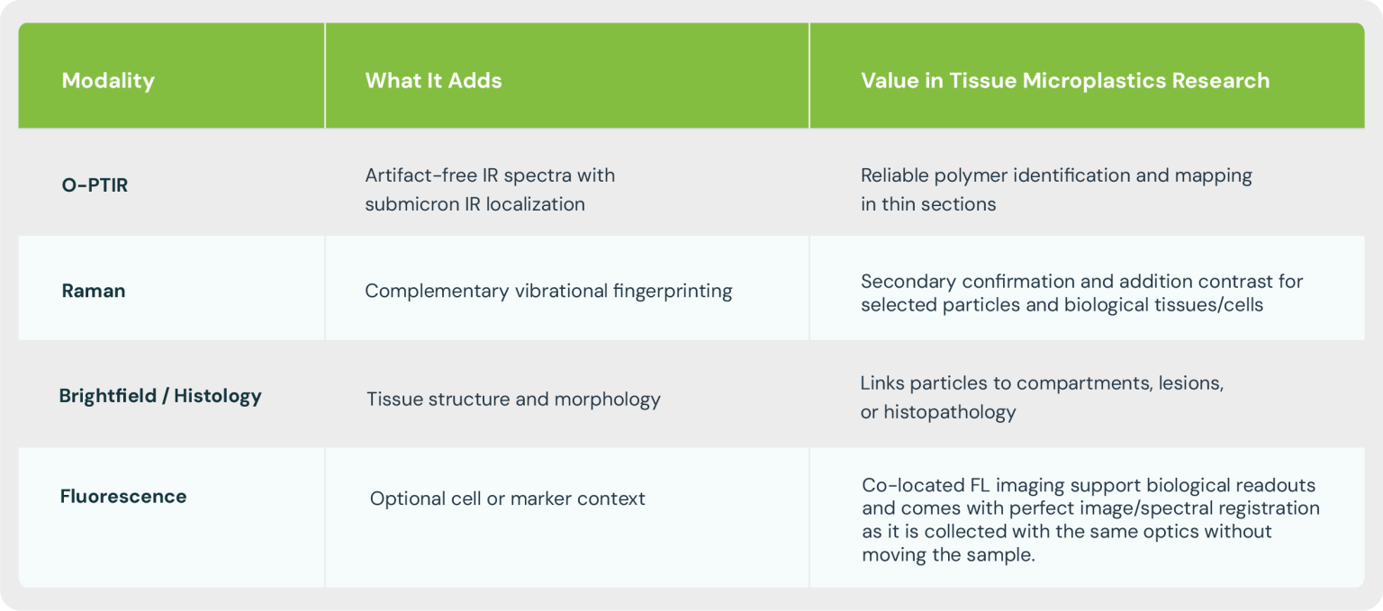
Multimodal tissue analytics: polymer ID plus biological context
O-PTIR becomes especially valuable when combined with simultaneous, co-located Raman capability and optional correlation with brightfield or fluorescence microscopy. This multimodal approach helps distinguish particle chemistry, cellular responses and allows the use traditional fluorescence labelling approaches.
Support for STOMP applicants
Explore our publications and webinars below to learn more about recent advances in microplastics analysis using photothermal IR spectroscopy.
With STOMP proposal deadlines approaching, early engagement is key. Connect with our team to discuss how mIRage™ can support your research and strengthen your proposal.
PSC works directly with researchers to strengthen STOMP proposals and enable submicron microplastics analysis.
- Access to mIRage systems: Through a growing network of partner laboratories
- Method development support: Guidance on workflows for submicron and nanoplastics
- Collaborative opportunities: Joint projects to advance measurement capabilities
- Application resources: Publications, webinars, and technical expertise
Talk to us about your STOMP proposal
Either email Jay Anderson or complete our contact us form for more information.
Publications
Method for label-free & non-destructive detection of microplastics in human formalin-fixed paraffin-embedded tissue sections
First Author: Elisabeth S. Gruber, Department of General Surgery, Medical University Vienna, Austria
Scientific Reports 15, 42637 (2025). doi: 10.1038/s41598-025-26751-1
Polystyrene nanoplastic exposure promotes amyloid misfolding and metabolic impairment at sub-lethal doses. A subcellular infrared imaging study
First Author: Iran Augusto Neves da Silva, Lund University, Sweden
Environmental Science: Nano 13, 1948–1961 (2026). doi: 10.1039/D5EN01181G
Label-free non-destructive spectroscopic detection of mixed microplastic uptake and differential effects on intestinal epithelial cells
First Author: Charlotte E. Sofield, University of Notre Dame Australia and Curtin University.
Journal of Hazardous Materials 504, 141283 (2026). doi: 10.1016/j.jhazmat.2026.141283
Label-free identification and imaging of microplastic and nanoplastic biouptake using optical photothermal infrared microspectroscopy.
First Author: Jun-Ray Macairan, Department of Chemical Engineering, McGill University
Environmental Science & Technology 59(30), 15612–15622 (2025). doi.doi.1021.est.4c14367
Advancing microplastic characterization in environmental samples using optical photothermal infrared (O-PTIR) spectroscopy
First Author: Moayad Yacoub, School of Earth, Environment, & Sustainability, Ball State University, USA
Environmental Geochemistry and Health 48, 279 (2026). doi.1007/s10653-026-03158-z
Optical photothermal infrared spectroscopy with simultaneously acquired Raman spectroscopy for two-dimensional microplastic identification
First Author: Julia Sophie Böke et al, Leibniz Institute of Photonic Technology
Scientific Reports 12, 18785 (2022). doi: 10.1038/s41598-022-23318-2
Webinars
- Life Science | Microplastics

- May 28, 2026
- Life Science | Microplastics
- April 1, 2026
- Life Science | Microplastics

- March 19, 2026
- Microplastics

- July 23, 2025
- Microplastics
- June 18, 2025
- Life Science | Microplastics

- June 26, 2025