
“Our findings show the potential of O-PTIR technology as a tool to study cancer on tissue samples… The principal advantage of O-PTIR over other IR imaging techniques such as FTIR is that it improves the spatial resolution from ~5-10 μm to ~0.5 μm yielding chemical information on a sub-cellular length scale.”
FULL PUBLICATION >
Reporting in Scientific Reports, researchers at the University of Liverpool utilized optical photothermal infrared (O-PTIR) spectroscopy combined with machine learning algorithms to evaluate 46 tissue cores of surgically resected cervical lymph nodes containing oral squamous cell carcinoma (OSCC) nodal metastases. The study aimed to determine whether O-PTIR could discriminate metastatic OSCC from normal lymphoid tissue with accuracy comparable to the gold standard of haematoxylin and eosin (H&E) stained histopathology. Traditional infrared spectroscopic techniques have been limited by spatial resolution of approximately 5-10 μm, making detailed cellular analysis challenging.
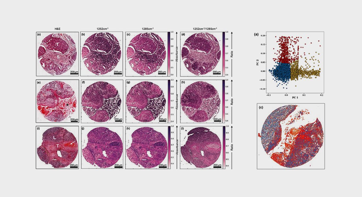
The research team collected O-PTIR single-wavenumber images at 1252 cm⁻¹, 1285 cm⁻¹, 1540 cm⁻¹, and 1660 cm⁻¹ from tissue microarrays. The ratio of O-PTIR chemical images at 1252 cm⁻¹ and 1285 cm⁻¹ successfully revealed morphological details comparable to H&E staining. When these ratio images were analyzed using a hybrid convolutional neural network (CNN) and random forest (RF) approach, the system achieved sensitivities of 98.6 ± 0.3%, specificities of 92 ± 4%, precision of 94 ± 5%, and an area under the receiver operator characteristic curve (AUC) of 94 ± 2%. Principal component analysis of the ratio images captured 79.7% of total variance and revealed three distinct tissue types: OSCC metastasis, lymphoid tissue, and stroma.
The enhanced spatial resolution of O-PTIR, approximately 0.5 μm compared to the 5-10 μm limitation of FTIR, enabled identification of cell nuclei and other sub-cellular structures critical for cancer morphology assessment. The technique successfully discriminated metastatic OSCC from surrounding tissues across samples with varying histological features, including those with heavy keratinization and different tumor-to-lymphoid tissue ratios. The ratio images showed superior tissue discrimination compared to single-wavenumber images, enhancing differences between OSCC and surrounding stroma while maintaining contrast similar to H&E images.
O-PTIR demonstrates significant potential as a label-free, high-resolution tool for cancer tissue analysis. The technique’s ability to provide chemical information at sub-cellular length scales while maintaining speeds practical for tissue analysis represents an important advancement over traditional FTIR imaging. The successful discrimination of metastatic oral cancer from normal tissue without staining, combined with high sensitivity and specificity, establishes O-PTIR as a promising technology for studying cancer tissue samples and potentially supporting clinical histopathology workflows.
Authors:
Safaa Al Jedani, Cassio Lima, Caroline I. Smith, Philip J. Gunning, Richard J. Shaw, Steve D. Barrett, Asterios Triantafyllou, Janet M. Risk, Royston Goodacre, Peter Weightman
Department of Physics, University of Liverpool
