
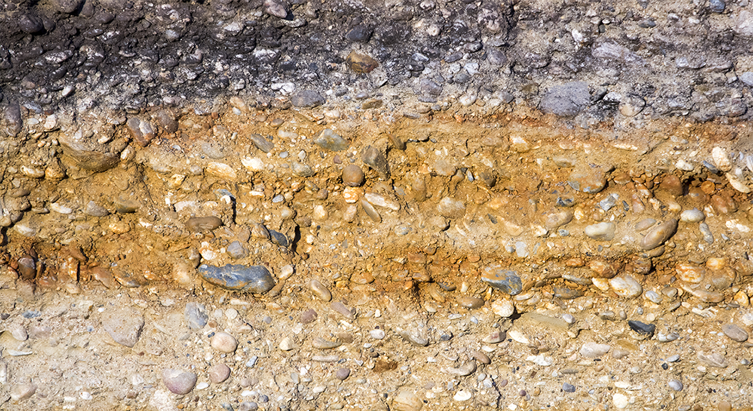
“O-PTIR microscopy opens the door to resolving the spatial association of microbes, minerals, and organic compounds down to the microscale and to tracking their interactions over time.”
FULL PUBLICATION >
Reporting in Environmental Science & Technology, researchers at the University of Lausanne, Switzerland, address a long-standing analytical challenge in soil and sediment science: resolving dynamic interactions among microbes, minerals, and organic matter at the microscale. Conventional Fourier-transform infrared (μ-FTIR) microscopy is limited to spatial resolutions of several micrometers and is susceptible to dispersive scatter artifacts, while the contact-based ATR-FTIR mode limits sample throughput and risks cross-contamination. These constraints have made real-time, nondestructive characterization of mineral-organic microstructures difficult to achieve.
The study systematically compared O-PTIR to ATR-FTIR and μ-FTIR across a range of environmentally relevant mineral and organic reference compounds. O-PTIR spectra of all minerals — including crystalline oxides (boehmite), phyllosilicates (kaolinite, montmorillonite), and poorly ordered phases (ferrihydrite) — showed spectral quality comparable to ATR-FTIR and substantially better than μ-FTIR, which showed almost no signal. O-PTIR detection sensitivity was greatest for highly crystalline minerals and for low molecular weight organic compounds. The authors note that variability in peak intensities observed for anisotropic phyllosilicates is attributable to differences in thermal expansion across crystal planes, and does not affect mineral identification.
To assess sensitivity toward mineral-bound organic matter, synthetic mineral-organic microstructures were prepared by coprecipitating glutamic acid with Al(OH)₃ across a range of carbon loadings. O-PTIR resolved recognizable absorption bands for both mineral and mineral-bound glutamic acid even at the lowest carbon concentration tested. The organic/inorganic signal ratio was consistently higher for O-PTIR than for ATR-FTIR across all carbon loadings, demonstrating greater sensitivity toward organic compounds — attributable to the higher thermal expansion capacity of organics. Using hyperspectral mapping at 2 μm step size over a 28 × 28 μm² area, O-PTIR further distinguished mineral-bound from unbound glutamic acid based on characteristic peak shifts associated with mineral interaction, resolving the chemical heterogeneity of the sample at the microscale.
O-PTIR microscopy opens the door to nondestructive, time-resolved analysis of microbe-mineral-organic matter interactions in soils and sediments. Operating without physical contact, free from diffuse and specular reflectance artifacts, and compatible with soil-on-a-chip and microfluidic systems, O-PTIR addresses an analytical gap that existing techniques — XRF, Raman microspectrometry, and NanoSIMS — cannot fill alone. The authors provide practical best-practice recommendations for damage-free acquisition, spectral interpretation of anisotropic compounds, and sample preparation for mineral-organic systems.
Authors:
Floriane Jamoteau, Mustafa Kansiz, Miriam Unger, and Marco Keiluweit
Institute of Earth Surface Dynamics, University of Lausanne, Switzerland
