
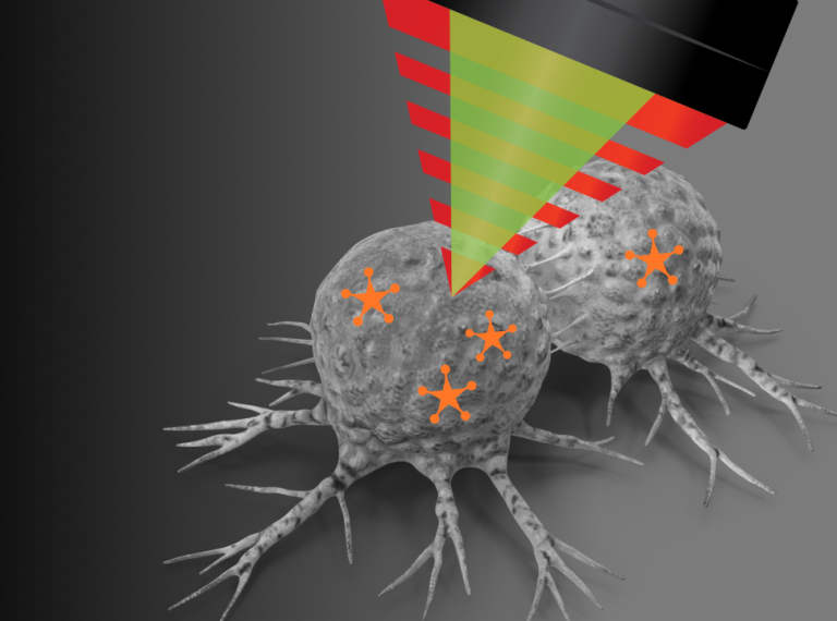
Vibrational tags & hybrid techniques
Bridge label-free and targeted imaging with bioorthogonal vibrational probes
The vibrational tags advantage
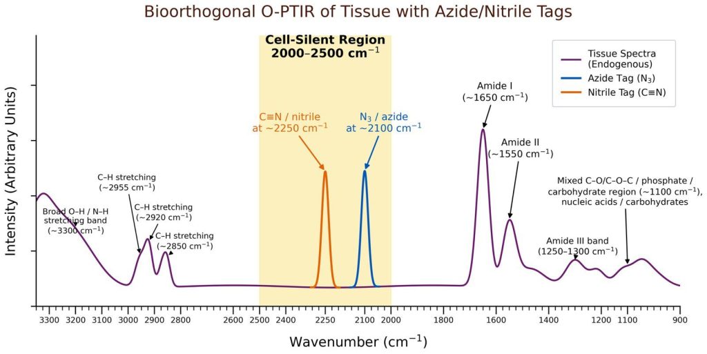
Vibrational tags exploit the “cell-silent region” (2800–1800cm⁻¹) of the IR/Ramanspectrum where endogenous biomolecules have minimal absorption. By incorporating bioorthogonal chemical tags—such as alkynes (C≡C), nitriles (C≡N), azides (N₃), and deuterium (²H)—researchers can track specific molecules with high sensitivity and no background interference.
O-PTIR’s submicron IR spatial resolution (~500 nm) combined with simultaneous Raman detection makes it ideal for:
- Direct detection of azide/nitrile-tagged biomolecules without click chemistry
- Correlation of vibrational tags with broad chemical imaging
- Multiplexed detection of multiple tags in complex samples
- Integration with FISH for function + identity mapping
- SERS enhancement for single-molecule sensitivity
~500 nm
Spatial resolution
2800–1800 cm⁻¹
Cell-Silent Region
Label-Free
Direct O-PTIR detection
Multimodal
IR + Raman + Fluorescence
Key technologies
Azide Tags (–N₃)
Azide groups are widely used bioorthogonal vibrational tags that produce strong infrared-active signals within the cell-silent region, making them highly attractive for direct O-PTIR detection workflows.
- Strong asymmetric N₃ stretch in the silent region (~2100 cm⁻¹)
- Excellent IR activity with minimal endogenous tissue interference
- Compatible with direct vibrational detection without secondary fluorescent labeling
- Small chemical handle with limited perturbation to native biology
- Suitable for multimodal IR, Raman, and fluorescence-correlative workflows
- Metabolic labeling of glycans and sugars
- Biomolecule trafficking studies
- Drug uptake and distribution
- Cellular metabolism investigations
- Correlative spatial biology workflows
Nitrile Tags (C≡N)
Nitrile groups provide sharp and spectrally isolated vibrational signatures within the cell-silent region, supporting chemically specific O-PTIR imaging with low biological background.
- Sharp C≡N stretch typically observed at ~2200–2300 cm⁻¹
- Strong spectral isolation from native biomolecular bands
- Compact bioorthogonal tag with minimal steric disruption
- Compatible with direct IR/O-PTIR vibrational detection
- Potential for multiplexed imaging using shifted nitrile frequencies
- Small-molecule and drug tracking
- Lipid and metabolite imaging
- Protein environment sensing
- Live-cell metabolic studies
- Multimodal chemical imaging workflows
Alkyne Tags (C≡C)
Alkyne groups produce sharp vibrational signatures within the cell-silent region (~2100–2200 cm⁻¹), making them widely used Raman tags and potentially compatible with direct O-PTIR detection without secondary click-label conjugation.
- C≡C stretch at ~2125 cm⁻¹ (EdU standard)
- No background interference from biomolecules
- Label nutrients, drugs, lipids, proteins
- Multiplexing with isotope editing (¹³C)
- DNA synthesis tracking (EdU)
- RNA transcription (5-ethynyl uridine)
- Protein synthesis (Hpg)
- Drug uptake & metabolism
- Lipid trafficking
Azide-Tagged FISH Probes
Fluorescence In Situ Hybridization (FISH) probes incorporating IR-active bioorthogonal tags such as azides may enable simultaneous molecular identification and chemically specific O-PTIR imaging within the same spatial workflow.
- Strong IR-active azide vibrational signature in the cell-silent region (~2100 cm⁻¹)
- Potential compatibility with direct O-PTIR detection without secondary reporter conjugatione
- Combines cellular identity with spatial chemical information
- Supports correlative fluorescence, IR, and Raman imaging workflows
- Potential multiplexing using orthogonal vibrational tags
- Microbial community analysis
- Spatial microbiome mapping
- Host–pathogen interaction studies
- Single-cell metabolic investigations
- Correlative spatial biology workflows
Raman-active
Raman-active vibrational tags such as alkynes may complement O-PTIR-FISH workflows by providing additional chemically specific contrast and multiplexing capabilities.
- Distinct vibrational signatures within the cell-silent region
- Compatible with correlative Raman and O-PTIR imaging
- Enables simultaneous molecular identity and metabolic analysis
- Supports isotope-labeling strategies
- Potential single-cell functional mapping
- Bacterial metabolism mapping
- Gene expression and chemical profiling
- Functional microbiology studies
- Spatially resolved metabolic imaging
- Multimodal microbial imaging workflows
SERS integration
Surface-Enhanced Raman Spectroscopy with O-PTIR enables concurrent SERS and SEIRA (Surface-Enhanced Infrared Absorption) for single-molecule detection.
- Enhancement factors up to 10¹⁴–10¹⁵
- Single-molecule sensitivity
- Concurrent SERS + SEIRA from O-PTIR
- Gold/silver nanoparticles or AFM tips
- Ultra-sensitive biosensing
- Single extracellular vesicle analysis
- Ultra-low concentration detection
- Antibody-conjugated SERS tags
- Protein biomarker detection
- Pharmaceutical analysis
The cell-silent region concept
Endogenous biomolecules have minimal IR/Raman absorption between 2800–1800 cm⁻¹, creating a “silent window” for bioorthogonal tags.
Research breakthroughs
Label-Free Single-Cell Chemistry
Submicron O-PTIR imaging enables chemical mapping at the cellular level with minimal sample prep, supporting high-confidence biomolecular identification.
- ~500 nm spatial resolution for subcellular features
- Simultaneous IR + Raman from the same location
- Reduced fluorescence/IR background vs. conventional methods
Metabolic Tracing in the Cell-Silent Region
Alkyne/isotope tagging supports direct detection in the 2100–2200 cm⁻¹ window, enabling clean metabolic readouts without click chemistry.
- Direct O-PTIR readout of C≡C vibrational signatures
- Minimal biomolecular background interference
- Multiplex-ready with isotope editing
Multimodal Identity + Function Mapping
Integrations like Raman-FISH and surface-enhancement approaches link “who is there” with “what they’re doing,” combining molecular identity with chemical phenotype.
- Species ID + metabolic activity in one workflow
- Compatible with isotope labeling and Raman tags
- High-sensitivity options via SERS/SEIRA-enhancement concepts
O-PTIR-FISH: Linking metabolism to identity
A groundbreaking protocol combining optical photothermal infrared imaging with fluorescence in situ hybridization (FISH) for simultaneous species identification and metabolic analysis at single-cell resolution.
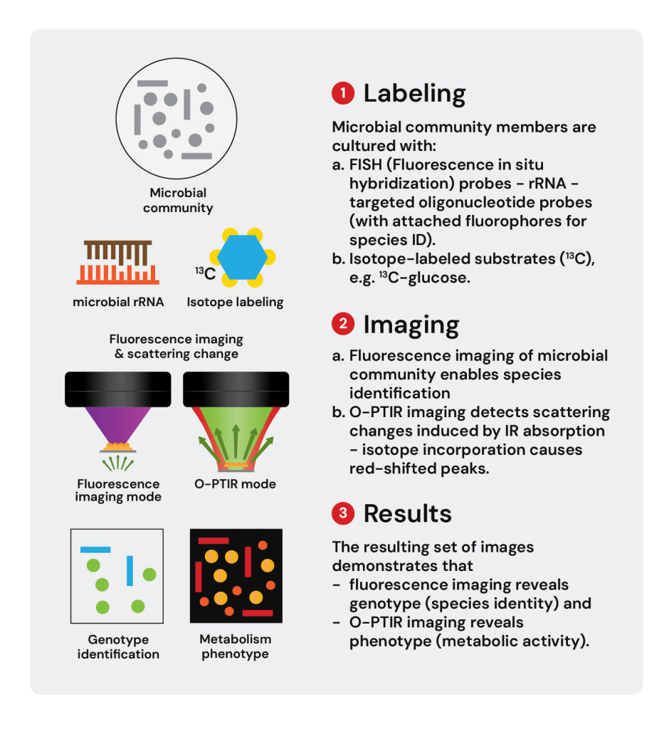
The O-PTIR-FISH workflow
This integrated platform addresses a critical challenge in microbiology: the concurrent task of differentiating cellular identity and elucidating metabolism at the single-cell level. Traditional methods like polymerase chain reaction and FISH allow species identification but lack metabolic information. O-PTIR-FISH solves this by combining:
- Isotope labeling – Culture with ¹³C-glucose or other labeled substrates
- FISH probes – rRNA-targeted oligonucleotides with fluorophores for species ID
- O-PTIR imaging – Detect isotope-induced red-shift in IR spectra
- Fluorescence imaging – Multi-channel fluorescence on same microscope
- Single-cell correlation – Link metabolism to taxonomic identity
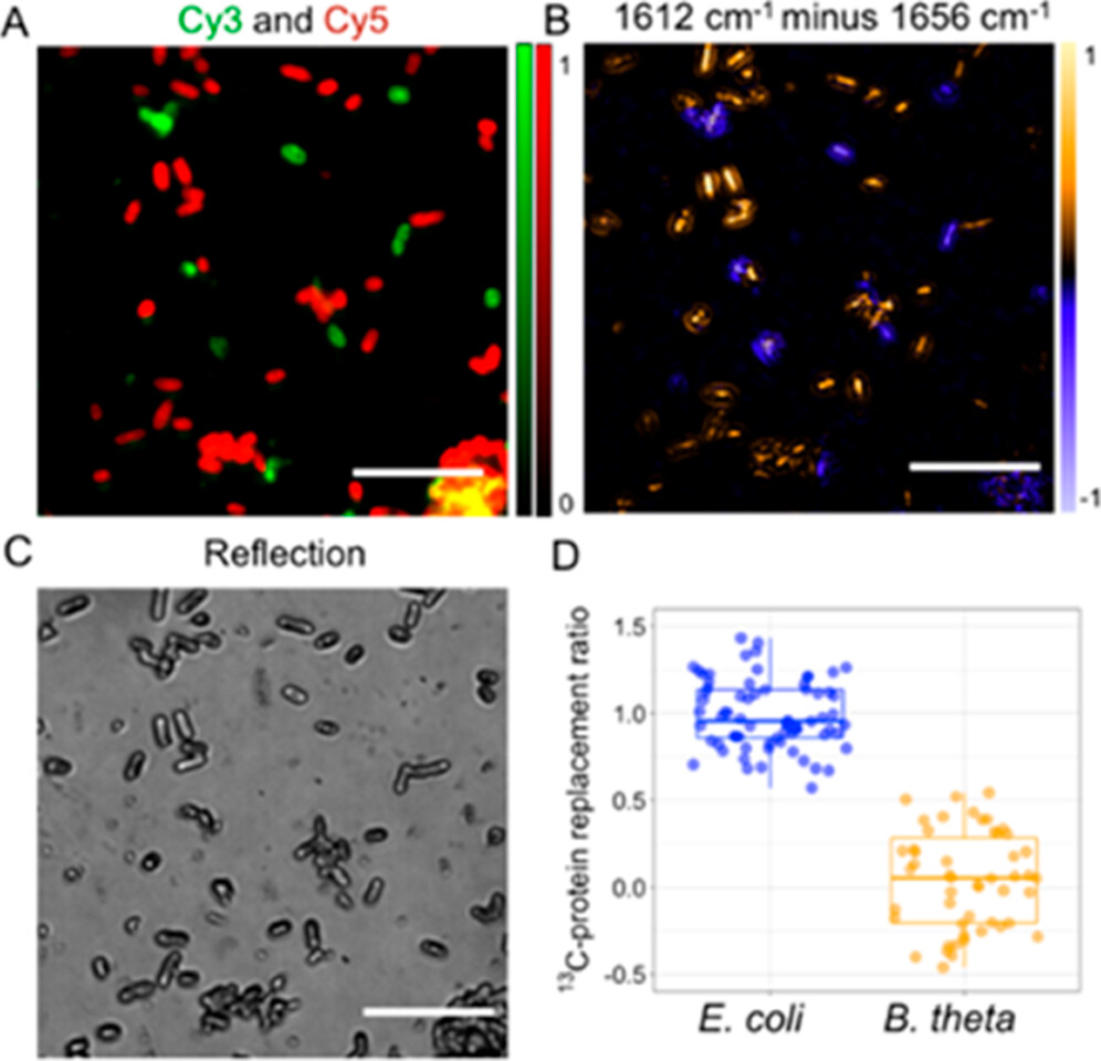
General workflow for single-cell analysis of metabolic activity with genetic identification
(A) Isotope labeling: Bacterial cells cultured with ¹³C-glucose in minimal medium. (B) FISH: Hybridization with fluorescent oligonucleotide probes targeting specific 16S rRNA sequences. (C) Sample preparation: 1-2 µL droplets on IR-transparent CaF₂ slide. (D) Bright-field imaging: Locate optimal cell density regions. (E) Multi-color FISH: Cy3 (green) and Cy5 (red) fluorophores reveal species identity (E. coli vs B. theta). (F-G) OPTIR imaging: At normal (1656 cm⁻¹) and shifted (1612 cm⁻¹) amide I peaks. Referencing unlabeled and fully labeled spectra (coefficients h), calculate isotopic replacement ratio = ci/(cr + ci). (H) Species ID: Multi-channel fluorescence identifies E. coli (Cy5), B. theta (Cy3), or unknown. (I) Correlation: Link metabolic activity (O-PTIR) to species identity (FISH) at single-cell level—enables high-throughput analysis in complex communities.
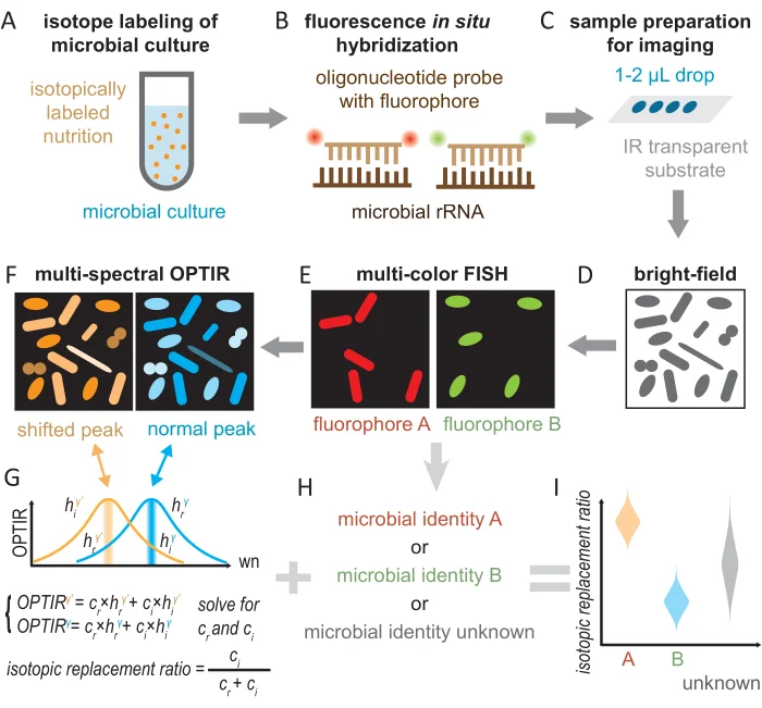
Isotope-induced spectral shift detection
The incorporation of heavier isotopes (¹³C) slows down chemical bond vibrations, leading to red-shifted peaks in vibrational spectra. For protein synthesis from ¹³C-glucose, the amide I peak red-shifts from 1656 cm⁻¹ (¹²C-protein) to 1612 cm⁻¹ (¹³C-protein)—a 44 cm⁻¹ shift easily resolved by O-PTIR.
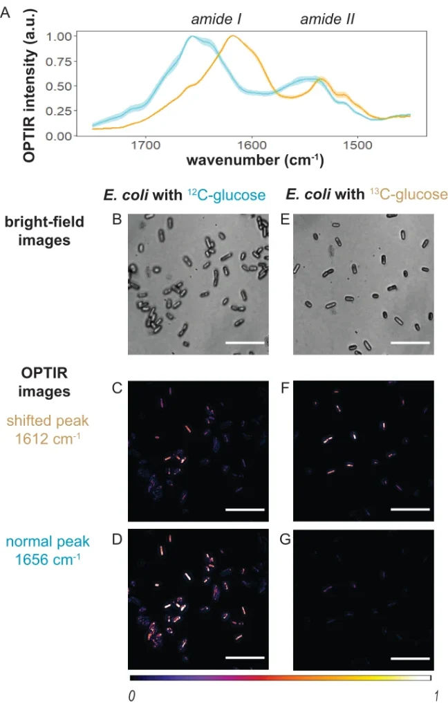
Representative O-PTIR spectra and imaging results from E. coli
(A) O-PTIR spectra: E. coli cultured with ¹²C-glucose (cyan) shows amide I peak at 1656 cm⁻¹ and amide II at ~1550 cm⁻¹. E. coli cultured with ¹³C-glucose (orange) shows red-shifted amide I at 1612 cm⁻¹ (44 cm⁻¹ shift). Shaded regions indicate standard deviation. (B-D) ¹²C-glucose E. coli: (B) Bright-field image shows rod-shaped bacteria. (C) O-PTIR at 1656 cm⁻¹ (normal peak) shows high intensity. (D) O-PTIR at 1612 cm⁻¹ (shifted peak) shows low intensity. (E-G) ¹³C-glucose E. coli: (E) Bright-field morphology. (F) Lower intensity at 1656 cm⁻¹. (G) Higher intensity at 1612 cm⁻¹, confirming ¹³C incorporation into proteins. Single cells are clearly resolved at ~500 nm resolution. Scale bars: 10 µm. This demonstrates O-PTIR’s ability to quantify metabolic incorporation of isotope-labeled substrates at the single-cell level.
Multi-species bacterial mixture analysis
To demonstrate O-PTIR-FISH’s capability in complex communities, researchers mixed two gut bacteria: E. coli (¹³C-labeled, Gam42a-Cy5 probe) and B. thetaiotaomicron (unlabeled, Bac303-Cy3 probe). The two species are morphologically similar (both rod-shaped), making microscopy alone insufficient for differentiation.
Representative Images and Quantification from Bacterial Mixtures
(A) Bright-field image: Mixture of E. coli and B. theta—both rod-shaped, indistinguishable by morphology alone. (B) Two-color FISH: E. coli labeled with Gam42a-Cy5 (red fluorescence). B. theta labeled with Bac303-Cy3 (green fluorescence). Clear species identification via rRNA-targeted probes. (C) O-PTIR subtraction (1612 cm⁻¹ − 1656 cm⁻¹): Positive pixels (orange/yellow) indicate ¹³C protein synthesis—these are E. coli cells cultured with ¹³C-glucose. Negative/blue pixels indicate ¹²C proteins—these are B. theta cultured with ¹²C-glucose. The subtraction map perfectly matches FISH identities. (D) Quantification: Isotopic replacement ratio (newly synthesized ¹³C-protein from ¹³C-glucose) shows statistically significant difference between species. E. coli: high ratio (orange distribution, mean ~0.8). B. theta: low ratio (blue distribution, mean ~0.2). Unknown cells: intermediate. Pairwise t-test: p = 9.74 × 10⁻³³. Scale bars: 10 µm. This demonstrates O-PTIR-FISH’s power to simultaneously identify species and quantify their differential metabolic activities in complex communities.
Key advantages of O-PTIR-FISH
Spatial Resolution
~500 nm resolution resolves individual bacterial cells and subcellular structures—comparable to fluorescence microscopy but with chemical specificity.
Single Setup
Both OPTIR and fluorescence imaging performed on same microscope—no complex alignment or co-registration needed between modalities.
Cultivation-Independent
FISH enables species identification without culturing—critical for non-culturable or slow-growing microbes in complex communities.
Quantitative Metabolism
Isotopic replacement ratio provides quantitative measure of metabolic incorporation—track kinetics, compare species, detect early changes.
High Throughput
Analyze hundreds of cells per field of view—enables statistical analysis of population heterogeneity and rare metabolic phenotypes.
Adaptable Probes
Extensive library of FISH probes available (probeBase database). Compatible with diverse isotopes (¹³C, ¹⁵N, ²H) and metabolic substrates.
“This protocol’s standardized and detailed nature will greatly facilitate its adoption by researchers from diverse backgrounds and disciplines within the broad single-cell metabolism research community.”
— Guo et al., JoVE (2024)
Advantages over traditional methods
| Feature | O-PTIR + Vibrational Tags | Fluorescence Imaging | Click Chemistry |
|---|---|---|---|
| Photobleaching | None | Major limitation | None (but requires azide-alkyne reaction) |
| Autofluorescence | No interference | High background in tissues | Depends on fluorophore |
| Chemical Signature | True vibrational fingerprint | Binary on/off signal | Requires reaction, fluorophore |
| Sample Preparation | Direct detection—no reaction | Staining protocols | Cu catalyst, washing steps |
| Spatial Resolution | ~500 nm (O-PTIR) | 200–300 nm (confocal) | Depends on imaging modality |
| Quantitative | Yes—peak intensity correlates with concentration | Semi-quantitative | Reaction efficiency variable |
| Broad Chemical Context | Simultaneous IR + Raman = full vibrational profile | No chemical info | Only tagged species |
| Live Cell Compatible | Yes—label-free readout | Yes | Requires fixation for click reaction |
Why O-PTIR for Vibrational Tags?
Cell replication studies
- S-phase DNA synthesis (EdU at 2125 cm⁻¹)
- Proliferation tracking in tumors
- Cell cycle analysis without click chemistry
- Tumor growth kinetics in tissue sections
- Alternative to BrdU antibody methods
Drug uptake & metabolism
- Azide-Tagged drugs for direct detection
- Cellular uptake kinetics
- Intracellular localization without fluorescence
- Metabolic conversion tracking
- Dose-response at single-cell level
Metabolic imaging
- Lipid synthesis (azido-palmitic acid)
- Protein synthesis (Hpg—alkyne methionine)
- Mitochondrial activity (MitoBADY)
- Multiplex: lipid + mitochondria simultaneously
Microbial communities
- O-PTIR-FISH: metabolism + species ID
- ¹³C-labeled and ¹5N-labelled substrate incorporation
- Spatial microbiome mapping
- Bacterial diversity in biofilms
- Host-microbe interactions
Organelle tracking
- Nucleus: EdU (DNA), EU (RNA)
- Mitochondria: BADY (2220 cm⁻¹, 25× EdU intensity)
- Lipid droplets: alkyne fatty acids
- Membrane lipid rafts: diyne-sphingomyelin
- Simultaneous multi-organelle imaging
Ultra-sensitive biosensing
- SERS + O-PTIR for single-molecule detection
- Protein biomarkers (10⁴–10⁵ enhancement)
- Extracellular vesicle analysis
- Antibody-conjugated SERS tags
- DNA/RNA sequence detection
Why O-PTIR for vibrational tags?
Submicron Spatial Resolution
~500 nm resolution resolves subcellular structures—no diffraction limit from mid-IR wavelengths. Probe beam defines resolution, not IR wavelength.
Direct Tag Detection
Detect Azide & Nitrile tags directly via O-PTIR—no click chemistry, no reaction artifacts, no fluorophore conjugation. True label-free readout after metabolic incorporation.
No Water Interference
O-PTIR’s photothermal detection overcomes water absorption that limits conventional FTIR in hydrated samples. Analyze fresh cells, tissues without drying.
Simultaneous IR + Raman
Same location, same time—correlate vibrational tag (Raman) with broad chemical composition (IR). Complementary info: IR strong for polar groups, Raman strong for C≡C/C=C.
Multimodal Integration
Combine with fluorescence (O-PTIR-FISH), SERS for enhancement, or isotope editing (¹³C, ²H) for multiplexing. Unparalleled chemical specificity.
Quantitative Analysis
Peak intensity correlates with tag concentration—track kinetics, dose-response, metabolic incorporation rates. No saturation effects from fluorescence quenching.
Ready to explore vibrational tags with O-PTIR?
Discover how alkyne tags, Raman-FISH, and SERS integration can transform your biomedical research with submicron chemical imaging.
Webinars
- Life Science | Microplastics

- May 28, 2026
- Life Science | Microplastics
- April 1, 2026
- Life Science
- February 12, 2026
- Life Science
- November 13, 2025
- Life Science

- October 2, 2025
- Life Science | Microplastics

- June 26, 2025
Need more information?
Discover how O-PTIR technology can elevate your research or help solve your toughest challenges. Our team are happy to assist and answer your questions.