
Advanced imaging techniques are at the heart of modern biomedical research, offering unparalleled insights into the structure and function of biological systems. Among these, multimodal imaging—integrating multiple complementary technologies—is a powerful approach to achieving greater specificity in biological analysis. One particularly promising combination is fluorescence (FL) imaging and infrared (IR) spectroscopy, a pairing that brings together the strengths of both methods for a more comprehensive understanding of complex biological samples.
By leveraging fluorescence’s ability to pinpoint specific molecular structures and infrared spectroscopy’s power to characterize chemical compositions, researchers can now obtain high-resolution, chemically rich images of biological tissues, cells, and biomolecules. This synergy is revolutionizing bioimaging applications, particularly in neurodegenerative disease research, where precise molecular characterization in precise locations is essential. Beyond this, multimodal imaging is also gaining traction in other fields, such as cancer research, cellular metabolism studies, and microbiology, expanding its impact across biomedical sciences.
What is Multimodal Imaging?
Multimodal imaging refers to the integration of two or more imaging techniques to overcome the limitations of individual modalities and provide a more complete view of biological processes. In the case of fluorescence and infrared spectroscopy:
- Fluorescence (FL) imaging provides high sptialspatial specificity, allowing researchers to target and visualize particular biomolecules, such as certain proteins, organelles or amyloid plaques. However, it lacks broad chemical information about the surrounding molecular environment.
- Infrared (IR) spectroscopy excels in chemical profiling, offering detailed insight into the composition of macromolecules, including proteins, lipids, and nucleic acids. However, it can lack some the specificity to single out particular molecular targets without additional guidance.
By integrating these two techniques, researchers gain both molecular specificity and chemical spatial resolution, allowing for more comprehensive biological insights. This approach is particularly beneficial in studying complex diseases like Alzheimer’s or Parkinson’s, where understanding molecular interactions at high resolution is essential.
How Combining FL and IR Creates a Powerful Tool
The fusion of fluorescence and infrared spectroscopy creates a robust platform for biological research by leveraging their complementary strengths:
- Targeting Specific Regions with FL Imaging: Fluorescent labelling allows scientists to identify and focus on particular structures, such as protein aggregates, cellular compartments, or biomarkers associated with diseases like Alzheimer’s.
- Gaining Chemical Insights with IR Spectroscopy: Once a region of interest is identified, IR spectroscopy provides detailed molecular characterization, allowing researchers to analyze the chemical environment in unprecedented detail or the molecular structures, for example, protein secondary structure. IR spectroscopy can also provide chemical images of the key distribution and relataive rations of macromolecules like proteins, lipids, nucleic acids and carbohydrates, all label-free.
- Enhanced Molecular Understanding: This dual approach is particularly suitable for the study of secondary protein structures, lipid distributions, and nucleic acid content, creating a more complete picture of biological samples.
- Reducing Potential Errors: Using a multimodal approach helps minimize data gaps or inconsistencies that could arise from relying on a single imaging method, leading to more robust and reproducible findings.
Such a combination is particularly valuable in studying protein misfolding diseases, where fluorescence can highlight amyloid aggregates and IR spectroscopy can analyze their structural composition.
Applications in Neurodegenerative Disease Research
One of the most critical applications of FL-IR multimodal imaging is in neurodegenerative disease research. Disorders such as Alzheimer’s and Parkinson’s are characterized by protein aggregation and structural misfolding, making them ideal candidates for this advanced imaging approach.
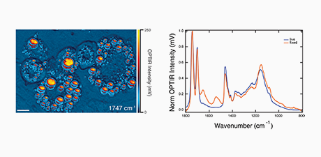
FL-OPTIR: Fluorescently Guided Optical Photothermal IR Microspectroscopy
A groundbreaking implementation of this approach is FL-OPTIR (fluorescently guided optical photothermal IR microspectroscopy). This technique:
- Allows researchers to pinpoint amyloid plaques and misfolded proteins with fluorescence markers.
- Uses IR spectroscopy to analyze the chemical structure and composition of these aggregates.
- Achieves sub-micron spatial resolution, enabling high-precision molecular studies of disease pathology.
Real-World Applications:
- Amyloid Plaques in Alzheimer’s Disease: Researchers have applied FL-OPTIR to examine the chemical composition of amyloid-beta plaques, gaining insights into their formation and structural properties, which is crucial for developing targeted therapies.
- Protein Misfolding in Parkinson’s Disease: The technique enables the detailed study of alpha-synuclein aggregation, aiding in early detection and characterization of disease progression.
By correlating fluorescence markers of disease pathology with infrared spectroscopic chemical analysis, scientists can more accurately study protein aggregation mechanisms, monitor disease progression, and assess therapeutic interventions.
Key Advantages of Multimodal Imaging
The combination of fluorescence and infrared spectroscopy offers numerous advantages that make it an indispensable tool in biomedical research:
- Increased Specificity: Fluorescence provides targeted visualization, allowing precise localization of disease-related biomolecules.
- Comprehensive Chemical Insights: IR spectroscopy offers detailed molecular characterization, complementing fluorescence’s structural specificity.
- Real-Time Imaging Possibilities: This technology holds the potential for real-time analysis in live-cell or live-tissue imaging, which is crucial for dynamic biological studies.
- Higher Resolution: Techniques like FL-OPTIR achieve sub-micron spatial resolution, surpassing the capabilities of traditional IR spectroscopy and fluorescence alone.
- Non-Destructive Analysis: Unlike some chemical staining techniques, FL-IR multimodal imaging preserves sample integrity, enabling longitudinal studies.
- Enhanced Reproducibility: By integrating complementary techniques, researchers can ensure more reliable, consistent data, reducing variability in experimental outcomes.
Conclusion
Integrating fluorescence and infrared spectroscopy represents a transformative step in bioimaging, offering unparalleled insights into the molecular landscape of biological systems. This multimodal approach is particularly powerful in neurodegenerative disease research, where understanding protein aggregation at both a structural and chemical level is crucial for developing effective therapies.
By combining the localization precision of fluorescence with the molecular depth of infrared spectroscopy, researchers can achieve a truly holistic view of biological systems. This cutting-edge technique is driving discoveries and shaping the future of biomedical imaging.
At Photothermal Spectroscopy Corp., we are committed to providing researchers with the most advanced multimodal imaging solutions to push the boundaries of bioimaging and disease research. Contact us today to learn how our technology can enhance your research in neurodegenerative diseases and beyond.
